Download

1 / 14
140 likes | 457 Vues
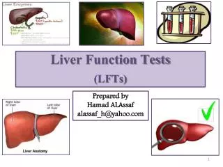
Liver function tests and functional tests of liver, ultrasound, biopsy. Liver Function Tests (LFT’s). These tests can be used to detect the presence of liver disease distinguish among different types of liver disorders gauge the extent of known liver damage follow the response to treatment.

E N D
Liver function tests and functional tests of liver, ultrasound, biopsy
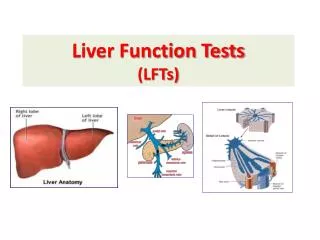
Liver Function Tests (LFT’s) These tests can be used to • detect the presence of liver disease • distinguish among different types of liver disorders • gauge the extent of known liver damage • follow the response to treatment. Liver tests have shortcomings • They can be normal in patients with serious liver disease and abnormal in patients with diseases that do not affect the liver • Liver tests rarely suggest a specific diagnosis; rather, they suggest a general category of liver disease, such as hepatocellular or cholestatic, and will help to decide whether the disease is acute or chronic
The Battery of Blood Tests • To increase both the sensitivity and the specificity of laboratory tests in the detection of liver disease, it is best to use them as a battery. • Those tests usually employed in clinical practice include: • bilirubin • Aminotransferases (ALT and AST) • alkaline phosphatase • Albumin • +/- prothrombin time • When more than one of these tests provide abnormal findings, or the findings are persistently abnormal on serial determinations, the probability of liver disease is high. • When all test results are normal, the probability of missing occult liver disease is low.
Our Patients Results • Albumin = 36 g/l (Ref range 35-45 g/l)Bilirubin(Total) = 38 umol/l (Ref range 0-17 umol/l)AlkP = 142 Units/l (Ref range 35-100 Units/l)GGT = 240 Units/l (Ref range 0-50 Units/l)ALT = 85 Units/l (Ref range 0-35 Units/l)AST = 160 Units/l (Ref range 0-42 Units/l) • Coagulation profile:Prothrombin time = 14 sec (Ref range 8-13s)APTT = 40 sec (Ref range 25-38s)Plasma Fibrinogen = 3.3 g/l (Ref range 1.5-4.0 g/L)
Whats What??? Enzymes that reflect damage to Hepatocytes: • Serum Aspartate aminotransferase (AST) • Serum alanine aminotransferase (ALT) • Serum lactate dehydrogenase (LDH) All cytosolic heptocellular enzymes. ie increase in blood indicates damage or death of hepatocytes ALT is the more specific to the liver The pattern of the aminotransferase elevation can be helpful diagnostically. In most acute hepatocellular disorders, the ALT is higher than or equal to the AST. An AST:ALT ratio > 2:1 is suggestive while a ratio > 3:1 is highly suggestive of alcoholic liver disease. The AST in alcoholic liver disease is rarely >300 U/L and the ALT is often normal. A low level of ALT in the serum is due to an alcohol-induced deficiency of pyridoxal phosphate.
Enzymes that Reflect damage to Bile Canaliculi: • alkaline phosphatase • 5'-nucleotidase • -glutamyl transpeptidase (GGT) Alkaline phosphatase can be elevated in many other conditions. GGT is not specific for cholestasis, also can reflect hepatocyte damage. In combination raised GGT can help determine if raised alkaline phosphatase elevations are due to liver disease Alkaline phosphatase and 5'-nucleotidase are found in or near the bile canalicular membrane of hepatocytes, while GGT is located in the endoplasmic reticulum and in bile duct epithelial cells. Reflecting its more diffuse localization in the liver, GGT elevation in serum is less specific for cholestasis than are elevations of alkaline phosphatase or 5'-nucleotidase.
Indicators of biliary excretory function: • Serum Bilirubin • Total (conjugated and unconjugated) • Direct: conjugated • Urine Bilirubin
Indicators of Hepatocyte Function: • Proteins secreted into blood • Serum albumin • Clotting factors ( prothrombin time) Serum albumin is synthesized exclusively by hepatocytes. Because of this slow turnover, the serum albumin is not a good indicator of acute or mild hepatic dysfunction; only minimal changes in the serum albumin are seen in acute liver conditions such as viral hepatitis, drug-related hepatoxicity, and obstructive jaundice. Hypoalbuminemia is more common in chronic liver disorders such as cirrhosis and usually reflects severe liver damage and decreased albumin synthesis. albumin levels < 3 g/dL should raise the possibility of chronic liver disease With the exception of factor VIII, the blood clotting factors are made exclusively in hepatocytes. Their serum half-lives are much shorter than albumin, ranging from 6 h for factor VII to 5 days for fibrinogen. Because of their rapid turnover, measurement of the clotting factors is the single best acute measure of hepatic synthetic function and helpful in both the diagnosis and assessing the prognosis of acute parenchymal liver disease. Useful for this purpose is the serum prothrombin time, which collectively measures factors II, V, VII, and X. Biosynthesis of factors II, VII, IX, and X depends on vitamin K. The prothrombin time may be elevated in hepatitis and cirrhosis as well as in disorders that lead to vitamin K deficiency such as obstructive jaundice or fat malabsorption of any kind.
Indicators of Hepatocyte Metabolism • Blood Ammonia • Ammonia is produced in the body during normal protein metabolism and by intestinal bacteria, primarily those in the colon. The liver plays a role in the detoxification of ammonia by converting it to urea, which is excreted by the kidneys. Striated muscle also plays a role in detoxification of ammonia, which is combined with glutamic acid to form glutamine. Patients with advanced liver disease typically have significant muscle wasting, which likely contributes to hyperammonemia in these patients.
Other laboratory tests • hepatitis serology to define the type of viral hepatitis • Reduced a1 antitrypsin levels, phenotypes PiZZ or PiSZ • Decreased serum ceruloplasmin and increased urinary copper; increased hepaticcopper level • Elevated iron saturation and serum ferritin; genetic testing for HFE gene mutations • autoimmune markers • to diagnose primary biliary cirrhosis (antimitochondrial antibody; AMA), sclerosing cholangitis (peripheral antineutrophil cytoplasmic antibody; P-ANCA), and autoimmune hepatitis (antinuclear, smooth-muscle, and liver-kidney microsomal antibody) • Serum Vitamin levels • not only in bile responsible for the absortption of fat-souble vitamins, the liver is respobsible for storage of all vitamins including water-soluble
Imaging • Ultrasound is the first diagnostic test to use. It shows • dilation of intrahepatic or extrahepatic biliary tree • Gallstones • Steatosis • space-occupying lesions within the liver (enables the clinician to distinguish between cystic and solid masses) • Doppler imaging can detect the patency of the portal vein, hepatic artery, and hepatic veins and determine the direction of blood flow • CT and MRI are indicated for: • identification and evaluation of hepatic masses • staging of liver tumours • preoperative assessment. • With regard to mass lesions, sensitivity and specificity remains a problem, and often two and sometimes three studies are needed before a diagnosis can be reached. Radiographic studies that strongly suggest cirrhosis include a small, nodular liver, ascites, splenomegaly, intra-abdominal varices, or portal and hepatic vein thrombosis; however, no test is considered a diagnostic gold standard. The current best test for diagnosing cirrhosis is liver biopsy.
Magnetic resonance cholangiopancreatography (MRCP) and endoscopic retrograde cholangiopancreatography (ERCP) are the procedures of choice for visualization of the biliary tree. (Through the endoscope, the physician can see the inside of the stomach and duodenum, and inject dyes into the ducts in the biliary tree and pancreas so they can be seen on xray) • Recently, methods using elastrography have been developed to measure hepatic stiffness as a means of assessing hepatic fibrosis. US elastrography is now undergoing evaluation for its ability to detect different degrees of hepatic fibrosis and to obviate the need for liver biopsy in assessing disease stage. • Nuclear Medicine Scans: show gallbladder filling and emptying. Inject radioactive dye, excreted by bile ie. accumulated in gallbladder, infusion of CCK, then can watch empty. • Interventional radiologic techniques allow the biopsy of solitary lesions, insertion of drains into hepatic abscesses, measurement of portal pressure, and creation of vascular shunts in patients with portal hypertension.
Percutaneous biopsy • There have been great advances made in hepatic imaging, although no method is suitably accurate in demonstrating underlying cirrhosis. Cirrhosis is identified by histopathologic examination of the liver • In selected instances, liver biopsy is necessary for diagnosis but is more often useful in assessing the severity (grade) and stage of liver damage, in predicting prognosis, and in monitoring response to treatment • Biopsy of the liver is a safe procedure that can be easily performed at the bedside with local anesthesia. Liver biopsy is of proven value in the following situations: (1) hepatocellular disease of uncertain cause (2) prolonged hepatitis with the possibility of chronic active hepatitis (3) unexplained hepatomegaly (4) unexplained splenomegaly (5) hepatic filling defects by radiologic imaging (6) fever of unknown origin (7) staging of malignant lymphoma. Liver biopsy is most accurate in disorders causing diffuse changes throughout the liver and is subject to sampling error in focal infiltrative disorders such as hepatic metastases. Liver biopsy should not be the initial procedure in the diagnosis of cholestasis.