Download
1 / 87
1.09k likes | 2.64k Vues
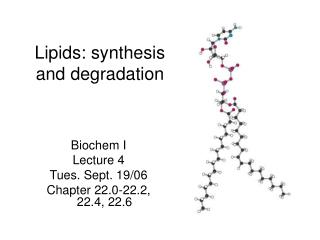
Amino Acid Degradation and Synthesis . UNIT IV: Nitrogen Metabolism. I. Overview . The catabolism of the amino acids found in proteins involves the removal of α-amino groups, followed by the breakdown of the resulting carbon skeletons.

E N D
Amino Acid Degradation and Synthesis UNIT IV: Nitrogen Metabolism
I. Overview • The catabolism of the amino acids found in proteins involves the removal of α-amino groups, followed by the breakdown of the resulting carbon skeletons. • These pathways converge to form seven intermediate products: oxaloacetate, α-ketoglutarate, pyruvate, fumarate, succinyl coenzyme A (CoA), acetyl CoA, and acetoacetate. • These products directly enter the pathways of intermediary metabolism, resulting either in the synthesis of glucose or lipid or in the production of energy through their oxidation to CO2 and water by the citric acid cycle.
Figure 20.1 Amino acid metabolism shown as a part of the central pathways of energy metabolism. (See Figure 8.2, p. 92, for a more detailed view of these processes.
Nonessential amino acids can be synthesized in sufficient amounts from the intermediates of metabolism or, as in the case of cysteine and tyrosine, from essential amino acids. • In contrast, the essential amino acids cannot be synthesized (or produced in sufficient amounts) by the body and, therefore, must be obtained from the diet in order for normal protein synthesis to occur. • Genetic defects in the pathways of amino acid metabolism can cause serious disease.
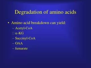
II. Glucogenic and Ketogenic Amino Acids • Amino acids can be classified as glucogenic, ketogenic, or both based on which of the seven intermediates are produced during their catabolism. A. Glucogenic amino acids • Amino acids whose catabolism yields pyruvate or one of the intermediates of the citric acid cycle are termed glucogenic or glycogenic. • These intermediates are substrates for gluconeogenesis and, therefore, can give rise to the net formation of glucose or glycogen in the liver and glycogen in the muscle.
B. Ketogenic amino acids • Amino acids whose catabolism yields either acetoacetate or one of its precursors (acetyl CoA or acetoacetyl CoA) are termed ketogenic. • Acetoacetate is one of the “ketone bodies,” which also include 3-hydroxybutyrate and acetone. • Leucine and lysine are the only exclusively ketogenic amino acids found in proteins. • Their carbon skeletons are not substrates for gluconeogenesis and, therefore, cannot give rise to the net formation of glucose or glycogen in the liver, or glycogen in the muscle.
III. Catabolism of the Carbon Skeletons of Amino Acids • The pathways by which amino acids are catabolized are conveniently organized according to which one (or more) of the seven intermediates listed above is produced from a particular amino acid. A. Amino acids that form oxaloacetate • Asparagine is hydrolyzed by asparaginase, liberating ammonia and aspartate (Figure 20.3). • Aspartate loses its amino group by transamination to form oxaloacetate (see Figure 20.3).
[Note: - Some rapidly dividing leukemic cells are unable to synthesize sufficient asparagine to support their growth. - This makes asparagine an essential amino acid for these cells, which therefore require asparagine from the blood. - Asparaginase, which hydrolyzes asparagine to aspartate, can be administered systemically to treat leukemic patients. - Asparaginase lowers the level of asparagine in the plasma and, therefore, deprives cancer cells of a required nutrient.]
B. Amino acids that form α-ketoglutarate 1. Glutamine is converted to glutamate and ammonia by the enzyme glutaminase. • Glutamate is converted to α-ketoglutarate by transamination, or through oxidative deamination by glutamate dehydrogenase. 2. Proline: This amino acid is oxidized to glutamate. • Glutamate is transaminated or oxidatively deaminated to form α-ketoglutarate. 3. Arginine: This amino acid is cleaved by arginase to produce ornithine. [Note: This reaction occurs primarily in the liver as part of the urea cycle.] • Ornithine is subsequently converted to α-ketoglutarate.
4. Histidine: This amino acid is oxidatively deaminated by histidase to urocanic acid, which subsequently forms N-formiminoglutamate (FIGlu, Figure 20.4). • FIGlu donates its formimino group to tetrahydrofolate, leaving glutamate, which is degraded as described above. • [Note: Individuals deficient in folic acid excrete increased amounts of FIGlu in the urine, particularly after ingestion of a large dose of histidine. The FIGlu excretion test has been used in diagnosing a deficiency of folic acid.]
C. Amino acids that form pyruvate 1. Alanine: This amino acid loses its amino group by transamination to form pyruvate. 2. Serine: This amino acid can be converted to glycine and N5,N10-methylenetetrahydrofolate. • Serine can also be converted to pyruvate by serine dehydratase (Figure 20.6B). • [Note: The role of tetrahydrofolate in the transfer of one-carbon units is presented on p. 267.] 3. Glycine: This amino acid can either be converted to serine by addition of a methylene group from N5,N10-methylenetetrahydrofolic acid (see Figure 20.6A), or oxidized to CO2 and NH3.
4. Cystine: This amino acid is reduced to cysteine, using NADH + H+ as a reductant. • Cysteine undergoes desulfuration to yield pyruvate. • [Note: The sulfate released can be used to synthesize 3'-phosphoadenosine-5'-phosphosulfate (PAPS), an activated sulfate donor to acceptors such as glycosaminoglycans.] 5. Threonine: This amino acid is converted to pyruvate or to α-ketobutyrate, which forms succinyl CoA.
Figure 20.6 A. Interconversion of serine and glycine, and oxidation of glycine. B. Dehydration of serine to form pyruvate.
D. Amino acids that form fumarate 1. Phenylalanine and tyrosine: Hydroxylation of phenylalanine leads to the formation of tyrosine (Figure 20.7). • This reaction, catalyzed by phenylalanine hydroxylase, is the first reaction in the catabolism of phenylalanine. • Thus, the metabolism of phenylalanine and tyrosine merge, leading ultimately to the formation of fumarate and acetoacetate. • Phenylalanine and tyrosine are, therefore, both glucogenic and ketogenic. 2. Inherited deficiencies: Inherited deficiencies in the enzymes of phenylalanine and tyrosine metabolism lead to the diseases phenylketonuria, and alkaptonuria, and the condition of albinism.
E. Amino acids that form succinyl CoA: methionine • Methionine is one of four amino acids that form succinyl CoA. • This sulfur-containing amino acid deserves special attention because it is converted to S-adenosylmethionine (SAM), the major methyl-group donor in one-carbon metabolism (Figure 20.8). • Methionine is also the source of homocysteine—a metabolite associated with atherosclerotic vascular disease.
Synthesis of SAM: Methionine condenses with adenosine triphosphate (ATP), forming SAM—a high-energy compound that is unusual in that it contains no phosphate. • The formation of SAM is driven, in effect, by hydrolysis of all three phosphate bonds in ATP (see Figure 20.8). 2. Activated methyl group: The methyl group attached to the tertiary sulfur in SAM is “activated,” and can be transferred to a variety of acceptor molecules, such as norepinephrine in the synthesis of epinephrine (see p. 286). • The methyl group is usually transferred to oxygen or nitrogen atoms, but sometimes to carbon atoms. • The reaction product, S-adenosylhomocysteine, is a simple thioether, analogous to methionine. • The resulting loss of free energy accompanying the reaction makes methyl transfer essentially irreversible.
3. Hydrolysis of SAM: After donation of the methyl group, S-adenosylhomocysteine is hydrolyzed to homocysteine and adenosine. • Homocysteine has two fates. If there is a deficiency of methionine, homocysteine may be remethylated to methionine. If methionine stores are adequate, homocysteine may enter the transsulfuration pathway, where it is converted to cysteine.
Resynthesis of methionine: • Homocysteine accepts a methyl group from N5-methyltetrahydrofolate (N5-methyl-THF) in a reaction requiring methylcobalamin, a coenzyme derived from vitamin B12. • The methyl group is transferred from the B12 derivative to homocysteine, and cobalamin is recharged from N5-methyl-THF.
b. Synthesis of cysteine: Homocysteine condenses with serine, forming cystathionine, which is hydrolyzed to α-ketobutyrate and cysteine (see Figure 20.8). • This vitamin B6–requiring sequence has the net effect of converting serine to cysteine, and homocysteine to α-ketobutyrate, which is oxidatively decarboxylated to form propionyl CoA. • Propionyl CoA is converted to succinyl CoA. • Because homocysteine is synthesized from the essential amino acid methionine, cysteine is not an essential amino acid as long as sufficient methionine is available.
4. Relationship of homocysteine to vascular disease: • Elevations in plasma homocysteine levels promote oxidative damage, inflammation, and endothelial dysfunction, and are an independent risk factor for occlusive vascular disease (Figure 20.9). • Mild elevations are seen in about 7% of the population. • Epidemiologic studies have shown that plasma homocysteine levels are inversely related to plasma levels of folate, B12, and B6—the three vitamins involved in the conversion of homocysteine to methionine or cysteine.
Supplementation with these vitamins has been shown to reduce circulating levels of homocysteine; however, the data do not prove the hypothesis that lowering homocysteine should result in reduced cardiovascular morbidity and mortality. • This raises the question as to whether homocysteine is a cause of the vascular damage or merely a marker of such damage. • [Note: Large elevations in plasma homocysteine as a result of rare deficiencies in cystathionine β-synthase are seen in patients with classic homocystinuria. • These individuals experience premature vascular disease, with about 25% dying from thrombotic complications before 30 years of age.]
Figure 20.9 Association between cardio-vascular disease mortality and total plasma homocysteine.
Figure 20.9 Effect of homocysteine-lowering therapy with folic acid, vitamin B12, and vitamin B6 on clinical outcome after coronary angioplasty. [Note: Balloon angioplasty is a noninvasive procedure in which a balloon-tipped catheter is introduced into a diseased blood vessel. As the balloon is inflated, the vessel opens further, allowing for placement of a stent and improved flow of blood.]
Elevated homocysteine levels in pregnant women are associated with increased incidence of neural tube defects (improper closure, as in spina bifida) in the fetus. • Periconceptual supplementation with folate reduces the risk of such defects.
F. Other amino acids that form succinyl CoA • Degradation of valine, isoleucine, and threonine also results in the production of succinyl CoA—a tricarboxylic acid (TCA) cycle intermediate and glucogenic compound. • Valine and isoleucine: These amino acids are branched-chain amino acids that generate propionyl CoA, which is converted to succinyl CoA by biotin- and vitamin B12–requiring reactions (Figure 20.10). • [Note: Propionyl CoA, then, is generated by the catabolism of certain amino acids and odd-numbered fatty acids (see p. 194).] 2. Threonine: This amino acid is dehydrated to α-ketobutyrate, which is converted to propionyl CoA and then to succinyl CoA. [Note: Threonine can also be converted to pyruvate.]
G. Amino acids that form acetyl CoA or acetoacetyl CoA • Leucine, isoleucine, lysine, and tryptophan form acetyl CoA or acetoacetyl CoA directly, without pyruvate serving as an intermediate (through the pyruvate dehydrogenase reaction). • As mentioned previously, phenylalanine and tyrosine also give rise to acetoacetate during their catabolism. Therefore, there are a total of six ketogenic amino acids. 1. Leucine: This amino acid is exclusively ketogenic in its catabolism, forming acetyl CoA and acetoacetate (see Figure 20.10). • The initial steps in the catabolism of leucine are similar to those of the other branched-chain amino acids, isoleucine and valine (see below).
2. Isoleucine: This amino acid is both ketogenic and glucogenic, because its metabolism yields acetyl CoA and propionyl CoA. • The first three steps in the metabolism of isoleucine are virtually identical to the initial steps in the degradation of the other branched-chain amino acids, valine and leucine (see Figure 20.10). 3. Lysine: An exclusively ketogenic amino acid, this amino acid is unusual in that neither of its amino groups undergoes transamination as the first step in catabolism. • Lysine is ultimately converted to acetoacetyl CoA. 4. Tryptophan: This amino acid is both glucogenic and ketogenic because its metabolism yields alanine and acetoacetyl CoA.
H. Catabolism of the branched-chain amino acids • The branched-chain amino acids, isoleucine, leucine, and valine, are essential amino acids. • In contrast to other amino acids, they are metabolized primarily by the peripheral tissues (particularly muscle), rather than by the liver. • Because these three amino acids have a similar route of catabolism, it is convenient to describe them as a group (see Figure 20.10).
Figure 20.10 Degradation of leucine, valine, and isoleucine. TPP = thiamine pyrophosphate.
1. Transamination: Removal of the amino groups of all three amino acids is catalyzed by a single, vitamin B6–requiring enzyme, branched-chain α-amino acid aminotransferase. 2. Oxidative decarboxylation: Removal of the carboxyl group of the α-keto acids derived from leucine, valine, and isoleucine is catalyzed by a single multienzyme complex, branched-chain α-keto acid dehydrogenase complex. • This complex uses thiamine pyrophosphate, lipoic acid, FAD, NAD+, and CoA as its coenzymes.
[Note: This reaction is similar to the conversion of pyruvate to acetyl CoA by pyruvate dehydrogenase (see p. 110) and the oxidation of α-ketoglutarate to succinyl CoA by α-ketoglutarate dehydrogenase (see p. 112).] • An inherited deficiency of branched-chain α-keto acid dehydrogenase results in accumulation of the branched-chain α-keto acid substrates in the urine. • Their sweet odor prompted the name maple syrup urine disease (see p. 272).
3. Dehydrogenation: Oxidation of the products formed in the above reaction yields α-β-unsaturated acyl CoA derivatives. • This reaction is analogous to the FAD-linked dehydrogenation described in the β-oxidation scheme of fatty acid degradation (see p. 192). 4. End products: The catabolism of isoleucine ultimately yields acetyl CoA and succinyl CoA, rendering it both ketogenic and glucogenic. • Valine yields succinyl CoA and is glucogenic. • Leucine is ketogenic, being metabolized to acetoacetate and acetyl CoA. • [Note: Branched-chain amino acid catabolism also results in glutamine and alanine being sent out into the blood from muscle.]
IV. Role of Folic Acid in Amino Acid Metabolism • Some synthetic pathways require the addition of single carbon groups. • These “one-carbon units” can exist in a variety of oxidation states. • These include methane, methanol, formaldehyde, formic acid, and carbonic acid. • It is possible to incorporate carbon units at each of these oxidation states, except methane, into other organic compounds. • These single carbon units can be transferred from carrier compounds such as tetrahydrofolic acid and S-adenosylmethionine to specific structures that are being synthesized or modified.
The “one-carbon pool” refers to single carbon units attached to this group of carriers. Note: • CO2, the dehydrated form of carbonic acid, is carried by the vitamin biotin, which is a prosthetic group for most carboxylation reactions, but is not considered a member of the one-carbon pool. • Defects in the ability to add or remove biotin from carboxylases result in multiple carboxylase deficiency; treatment is supplementation with biotin.
A. Folic acid: a carrier of one-carbon units • The active form of folic acid, tetrahydrofolic acid (THF), is produced from folate by dihydrofolate reductase in a two-step reaction requiring two moles of NADPH. • The carbon unit carried by THF is bound to nitrogen N5 or N10, or to both N5 and N10. • THF allows one-carbon compounds to be recognized and manipulated by biosynthetic enzymes. • Figure 20.11 shows the structures of the various members of the THF family and their interconversions, and indicates the sources of the one-carbon units and the synthetic reactions in which the specific members participate. • [Note: Folate deficiency presents as a megaloblastic anemia due to decreased availability of the TMP needed for DNA synthesis (see p. 303).]
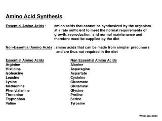
V. Biosynthesis of Nonessential Amino Acids • Nonessential amino acids are synthesized from intermediates of metabolism or, as in the case of tyrosine and cysteine, from the essential amino acids phenylalanine and methionine, respectively. • The synthetic reactions for the nonessential amino acids are described below, and are summarized later in Figure 20.14. • [Note: Some amino acids found in proteins, such as hydroxyproline and hydroxylysine, are modified after their incorporation into the protein (posttranslational modification]
Figure 20.11 Summary of the interconversions and uses of the carrier, tetra-hydrofolate.
A. Synthesis from α-keto acids • Alanine, aspartate, and glutamate are synthesized by transfer of an amino group to the α-keto acids pyruvate, oxaloacetate, and α-ketoglutarate, respectively. • These transamination reactions (Figure 20.12, and see p. 250) are the most direct of the biosynthetic pathways. • Glutamate is unusual in that it can also be synthesized by the reverse of oxidative deamination, catalyzed by glutamate dehydrogenase (see p. 252).
B. Synthesis by amidation • Glutamine: • This amino acid, which contains an amide linkage with ammonia at the γ-carboxyl, is formed from glutamate by glutamine synthetase. • The reaction is driven by the hydrolysis of ATP. • In addition to producing glutamine for protein synthesis, the reaction also serves as a major mechanism for the detoxification of ammonia in brain and liver.
2. Asparagine: • This amino acid, which contains an amide linkage with ammonia at the β-carboxyl, is formed from aspartate by asparagine synthetase, using glutamine as the amide donor. • The reaction requires ATP, and, like the synthesis of glutamine, has an equilibrium far in the direction of asparagine synthesis.
Figure 20.12 Formation of alanine, aspartate, and glutamate from the corresponding α-keto acids.
C. Proline • Glutamate is converted to proline by cyclization and reduction reactions. D. Serine, glycine, and cysteine • Serine: This amino acid arises from 3-phosphoglycerate, an intermediate in glycolysis, which is first oxidized to 3-phosphopyruvate, and then transaminated to 3-phosphoserine. • Serine is formed by hydrolysis of the phosphate ester. • Serine can also be formed from glycine through transfer of a hydroxymethyl group by serine hydroxymethyl transferase.
2. Glycine: This amino acid is synthesized from serine by removal of a hydroxymethyl group, also by serine hydroxymethyl transferase. 3. Cysteine: This amino acid is synthesized by two consecutive reactions in which homocysteine combines with serine, forming cystathionine, which, in turn, is hydrolyzed to α-ketobutyrate and cysteine (see Figure 20.8). • Homocysteine is derived from methionine as described on p. 264. • Because methionine is an essential amino acid, cysteine synthesis can be sustained only if the dietary intake of methionine is adequate.