Download
1 / 57
600 likes | 993 Vues
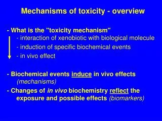
Mechanisms for toxicity of protein oxidation products. Oxidation of macromolecules lipids, proteins and DNA, produces various oxidation end products. These end products can be measured to assess oxidative stress in vivo. . Protein Oxidation.

E N D
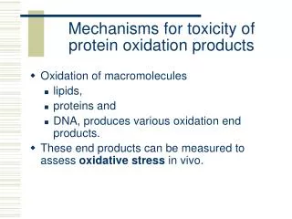
Mechanisms for toxicity of protein oxidation products • Oxidation of macromolecules • lipids, • proteins and • DNA, produces various oxidation end products. • These end products can be measured to assess oxidative stress in vivo.
Protein Oxidation • Proteins are continuously oxidized, even under normal physiological conditions. • This oxidation may increase in various disorders • including atherosclerosis, Parkinson’s disease and aging. • All amino acid residues of a protein are subject to radical attacks by reactive oxygen and nitrogen species; however, • Tyr, Phe, Trp, His, Met, and Cys residues are the preferred target sites for hydroxyl radicals.
Oxidized amino acids • Until now, oxidized amino acids have primarily been considered as molecular fingerprints (biomarkers) of oxidized protein formation. • Neither the toxic effects of these modified amino acids nor their metabolic fates have been investigated.
Questions • Q1. What are these oxidized amino acids? • Structures of OAA • Origins of OAA • Q2. Are these protein oxidation byproducts (OAA) toxic? • In vitro toxicity assays • Chinese hamster ovary cells • PC-12 cells (neuronal cell line) • In vivo model • Phenylketonuria • Animal study
More questions • Q3. What is the underlying biochemical mechanism of their toxicity? • might be incorporated into cellular proteins and disrupt their normal function? • Therefore, this study focuses on investigation of the metabolic fates of OAA, particularly • ortho-tyrosine, • meta-tyrosine, • o,o'-dityrosine, • 3-chlorotyrosine and • 3-nitrotyrosine in in vitro and in vivo systems.
i) The tyrosyl radical pathway (di-Tyr). • Photo- or chemical oxidation of L-tyrosine generates the long-lived tyrosyl radical which has been detected by electron paramagnetic resonance spectrometry. • When two tyrosyl radicals productively interact, the major product is o,o'-dityrosine, an intensely fluorescent compound. • Heinecke and others have shown that the tissue levels of protein-bound dityrosine increase in a wide variety of conditions associated with oxidative stress in vivo, including atherosclerosis, Parkinson's disease, and Alzheimer's disease.
ii) The myeloperoxidase pathway (3Cl-Tyr). • One well-characterized pathway for generating oxidants involves the NADPH oxidase of phagocytes, which produces superoxide (O2-). The O2- then dismutates into hydrogen peroxide (H2O2), an oxidizing substrate for myeloperoxidase, a heme protein secreted by activated phagocytes. O2- + O2- + 2H+ H2O2 + O2 • Myeloperoxidase uses H2O2 generated by this system to produce potent toxins. Myeloperoxidase is the only human enzyme known to generate HOCl. • Heinecke et al. recently demonstrated that myeloperoxidase converts tyrosine into 3-chlorotyrosine, a stable product that may serve as a molecular fingerprint of the enzyme's action.
iii)The metal ions and hydroxyl radical pathway (o-Tyr and m-Tyr). • Aromatic amino acids are important targets for metal ion-catalyzed damage. • Phenylalanine readily undergoes a radical addition reaction, yielding o-tyrosine and m-tyrosine. • Large amounts of o-tyrosine and m-tyrosine are generated in proteins oxidized by H2O2 and copper, a classic hydroxyl radical-generating system.
iv) The reactive nitrogen pathway (3-nitro-Tyr). • NO is a relatively stable free radical produced by nitric oxide synthase expressed in endothelial and other cells. • In vitro, NO reacts with superoxide to produce peroxynitrite (ONOO-): • NO + O2•- ONOO- + H+ ONOOH • In vitro studies demonstrate that ONOO (or species derived from ONOO such as ONOOH) promotes oxidation of proteins, nucleic acids and lipids and it is a potent cytotoxin. Among the protein oxidation products formed by ONOO is 3-nitrotyrosine.
Overall goal • Therefore, the overall goal of this study is to understand the metabolism of oxidized amino acids by using in vivo and in vitro models.
In vitro toxicity assays • CHO cells • Colony formation • Survival Fractions • Survival curves • LDH assay • 24 h incubation • 48 h incubation • MTS assay
Colony Formation Assays • Cytotoxicity of CHO cells was analyzed by a colony formation assay. Exponentially growing cells were collected after trypsinization and centrifuged at 1000xg for 5 min. • Between 100-2000 cells were plated into small (60 mm) petri dishes • Cells were then exposed to OAA for 7-10 days. • Following this incubation period, the resulting cell colonies were stained with methylene blue and counted.
Colony formation assay CONTROL Incubated with antibiotic degradation byproduct
Surviving fractions • The colony efficiency (CE) was calculated as: Colonies counted/ Cells seeded x100. • A cell survival curve was constructed by plotting the surviving fraction (number of colonies counted divided by the number of cells seeded times the colony efficiency of the control) from the groups versus OAA concentrations.
Colony formation results • Incubation of CHO cells with three OAA (meta, ortho and 3-nitrotyrosine) inhibited colony formation in a concentration dependent manner. • At 0.2 mM concentration of the meta-tyr, survival fraction was 62 % as compared to 98% of tyrosine at the same concentration.
Lactate dehydrogenase (LDH) assay • LDH is an intracellular enzyme. • LDH activity in the media is often used as a membrane damage marker. • CHO cells were incubated with ortho-tyrosine, meta-tyrosine and phenylalanine for 24 and 48 hours. The media was then analyzed for LDH activity based on the fact that LDH catalyzes the interconversion of lactate and pyruvate. • Pyruvate + NADH + H ---------- Lactate + NAD • In the presence of LDH, NADH is oxidized to NAD, resulting in a reduction in the absorbance at 340 nm.
Groups LDH (U/L) 24 hrs LDH (U/L) 48 hrs Control Phe or Tyr (5 mM) 44±0 43±0 48±2 43±0 m-Tyr (5 mM) 354±36 455±21 o-Tyr (5 mM) 76±3 434±0 LDH levels in the media
LDH results • LDH increased significantly in the presence of m-tyr and o-tyr, consistent with membrane damage.
MTS assay in CHO cells • Viability of CHO cells was assessed by using a 3-(4,5-dimethylthiazol-2-yl)-5-(3-carboxymethoxyphenyl)-2-(4-sulfophenyl)-2H-tetrazolium (MTS) assay. • The MTS tetrazolium compound is reduced by cells into a colored formazan product. • The absorbance at 490 nm is directly proportional to number of living cells.
CHO cells viability decreases when cells are incubated with meta-tyrosine.
Neurotoxicity of OAA • Two neurotoxicity assays were performed by using a pheochromacytoma cell line called PC-12. • MTS assay • Neurite outgrowth assay • PC-12 cells form neurites upon exposure to a nerve growth factor. • Any compound inhibiting the number of neurites per cell, or the length of each neurite, is said to be neurotoxic.
Both meta-tyrosine and ortho-tyrosine inhibited a cell's neurite-forming ability.
Cells completely lost their neurites with 1 mM ortho-tyrosine. Control 1mM p-Tyr 1mM o-Tyr 0.5 mM o-Tyr
Therefore • OAA were proven to be TOXIC by • Clonagenic assay • LDH assay • MTS assay • OAA were also shown to be neurotoxic by • MTS assay • PC-12 neurite outgrowth assay.
Q: Why are OAA toxic and neurotoxic? • In order to understand the underlying mechanism of observed cytotoxicity and neurotoxicity of OAA, their possible incorporation into proteins was investigated in in vitro and in vivo systems.
Aminoacylation / Translation Pathway OAA A C C 3’ A C C 3’ Aminoacyl t-RNA 5’ 5’ OAA + synthetase mischarged t-RNA t-RNAphe Translation (or t-RNAtyr) OAA OAA OAA OAA-incorporated protein OAA OAA Possible Degradation Pathways Amino acids peptides
Synthesis of radiolabeled amino acids • All of the necessary synthetic methods were routinely prepared in the Heinecke laboratory at Washington University. • 3-Nitro[14C]tyrosine is synthesized using L-[14C]tyrosine and tetranitromethane under basic conditions. • Ortho-[14C]tyrosine and meta-[14C]tyrosine are prepared from L-[14C]phenylalanine using hydroxyl radical generated by copper sulfate and H2O2 . Oxidation products are purified by reverse-phase HPLC.
Incorporation of OAA into proteins in CHO cells • CHO cells were plated at a density of 4x10⁵ cells/well in 6-well plates. • 14C-labeled tyrosine, phenylalanine, m-tyrosine and o-tyrosine were then added to the media for different groups and incubations were started. At the end of the indicated incubation times (6, 12, 24 hours), cells were trypsinized and harvested. Following immediate centrifugation, 20% TCA was added to the cell pellets and left on ice for 30 minutes for protein precipitation. • Proteins were pelleted by centrifuging the cells at 1000 xg at 4C for 4 min. The protein pellets were then washed two times with ice-cold saline.
Then, • To measure the amount of radioactivity in the proteins: the proteins were solubilized in 2% Triton X-100. An aliquot of the solubilized proteins was mixed with a scintillation mixture and the radioactivity of each group was counted in the liquid scintillation counter.
The incorporation of 14C-labeled tyr, m-tyr and o-tyr into proteins of CHO cells
Incorporation of m-tyr and o-tyr (Control cells: plain medium and labeled amino acids)
Identification of incorporated OAAs by HPLC • Incorporated OAA was further identified by acid hydrolysis of the cells that were incubated with 14C-labeled tyr, meta-tyrosine, and ortho-tyrosine for 24 hours. After hydrolysis of the proteins, the amino acids were injected onto a HPLC column for separation. HPLC fractions were collected and counted in a liquid scintillation counter. • Following figure shows both HPLC chromatograms from the separation of the acid hydrolysate samples, with cold amino acids included, and overlapped with a graph of counts for each fraction obtained with a scintillation counter
RT for para-Tyr is 8.43 min RT for m-Tyr is 9.83 min RT for o-tyr is 12.96 min
In vitro transcription/translation • The incorporation of m-tyrosine and o-tyrosine into luciferase during in vitro coupled transcription/translation is studied in a TNT® SP6 Coupled Reticulocyte Lysate System. • Luciferase SP6 Control DNA was incubated with 14C-labeled m-tyrosine or o-tyrosine in the presence of various amino acid compositions, RNA polymerase, rabbit reticulocyte lysate and RNasin ribonuclease inhibitor at 30 C for 60 min. 14C-labeled Tyr and Phe were used as positive controls for incorporation. Possible incorporation of OAA into luciferase was examined by gel electrophoresis.
SDS-PAGE and visualisation of radiolabeled proteins • Sodium dodecyl sulfate-polyacrylamide gel electrophoresis (SDS-PAGE) of translated luciferase was carried out in a Bio-Rad Mini-PROTEAN II cell. Samples were applied to gels after being diluted with loading buffer [-mercapthoethanol and a Laemmli sample buffer 1:19 v/v], at a ratio of 1:1 (v/v). A 25mM Tris, 192mM glycine and 0.1% (w/v) SDS mixture (pH 8.3) was used as a running buffer. Then samples were electrophoresed at a constant current of 130V per gel rod.
14C-labeled protein was visualized as follows; • After electrophoresis, proteins were fixed by placing the gel in 10% glacial acetic acid + 30% methanol mixture for 1 h. Then the gel was treated with autoradiography enhancer for an hour and then in cold water for 30 min. Then the gels were dried under vacuum at 60 C and exposed to X-ray film at –80 C for at least 2 weeks.
OAA incorporation • “Amino acid mixture” includes 20 amino acids at 1 mM concentration. As seen on lane D, translation in the presence of the amino acid lacking Phe resulted in incorporation of 14C-labeled m-tyr into luciferase. O-tyr was not found to be incorporated into luciferase in any of the translation conditions mentioned above.
A) without cold Phe + 14C-phe. B) without cold Tyr + 14C-tyr. C) without cold Tyr + 14C-m-tyr. D) without cold Phe + 14C-m-tyr. E) without cold Tyr + 14C-o-tyr. F) without cold Phe + 14C-o-tyr.
So, OAA are incorporated! • CHO cell experiments • In vitro translation experiments • Visualization by SDS-PAGE • All showed that OAA were incorporated into proteins.
In vivo model “PKU model” • We used a phenylketonuria (PKU) animal model as our in vivo model where Phe levels were very high due to Phe hydroxylase deficiency. • It has been shown that Phe could be used as a substrate for another enzyme, tyrosine hydroxylase, that produces meta-tyrosine and ortho-tyr, by its action on Phe. • Therefore, the presence of protein bound meta- and ortho-tyrosine in PKU could be expected in such an in vivo system if their incorporation into proteins occurred.
Hypothesis PAH Phe Phe Phe Phe Tyr Tyr Tyr Tyr Tyr hydroxylase or Nonspecific hydroxylation Enzymes • o-tyr • M-tyr • ??? • Phe acetate • Phe pyruvate • Phe lactate Damage to Neurons?
about PKU • PAH is on chromosome 1 • Population genetics • 1 in 10,000 • Metabolic phenotypes and mechanisms of neurotoxicity • The major clinical effect is on brain development and function • Neurotoxicity • Phe--------> is the chief villian?????
Neurochemistry of PKU • High plasma Phe values are associated with measurable acute impairment of higher brain functions • Abnormal EEG • High Phe levels alter brain chemistry • These acute effects are observed when plasma Phe is higher than 1300 uM (normal level: ~60 uM) • What causes neurotoxicity in PKU? • Decrease in Tyr? • Phe metabolites (Phe acetate, Phe pyruvate, and Phe lactate)? • No single mechanism seems to be responsible!
More about PKU • The cause of defective brain myelination in PKU has long been a focus of interest. • Brain protein synthesis affected by high Phe • Initiation of protein synthesis is inhibited. • High Phe causes: • Increase in loss of neurons in the rodent. • DNA content is decreased in affected brain cells • Net effect: IMPAIRED BRAIN GROWTH • Phe ITSELF IS PERHAPS THE NEUROTOXIC AGENT IN PKU or • o- and m-tyr??
PKU animals • ENU mice as a PKU animal model: Wild-type mice (RTBR/Pas background) were treated with an alkylating agent (N-ethyl-N’-nitrosourea: ENU) to induce mutations on the phenylalanine hydroxylase locus (PAH). Twelve animals (6 ENU1/1 as control, 6 ENU2/2 as PKU animal model), ranging in age from 14 to 24 weeks old, were sacrificed under anesthesia. Brain samples were dissected, immediately placed in ice-cold antioxidant buffer [50mM NaHPO4, pH 7.4, 100M diethylenetriaminepentaacetic acid, 1mM butylated hydroxytoluene, 1% (v/v) ethanol] and kept at –70 0C until analysis. This experiment was repeated at least three times.