

Abb26.19
E N D
Presentation Transcript
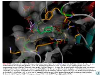
Abb. 26.19Kristallographisch ermittelter Bindungsmodus eines phosphorylierten Tyrosins (26.39, grün, Abb. 26.21) als minimales Mimetikum für ein Peptidsubstrat in der humanen Phosphatase PTP-1B. Die Phosphatgruppe wird durch einen Argininrest (221) in Position gehalten und auf den nucleophilen Angriff durch Cys 215 vorbereitet. Oberhalb des Cys-Rests ist Asp 181 zu finden, das für den Ausgleich der Protonen sorgt. Der Eingang der Bindetasche wird durch die beiden aromatischen Reste Phe 182 und Tyr 46 begrenzt. Die Bindestelle des Cysteins befindet sich am Ende einer langen Helix. Die gezeigte Geometrie beruht auf einer Kristallstruktur mit der katalytisch praktisch unwirksamen Cys→Ser-Mutante. In der Kristallstruktur ist ein zweites Phosphotyrosin (rosa) zu finden, das an Arg 24 und Arg 254 in einer zweiten distalen Tasche bindet. Die Besetzung dieser zweiten Bindetasche hat im Folgenden die Entwicklung nanomolarer Inhibitoren für die PTP-1B geprägt (vgl. Abb. 26.20).