
Schistosomiasis-Induced Longitudinally Extensive Transverse Myelitis: Case Reports and Insights
This study highlights three cases of schistosomal myelitis presenting as longitudinally extensive transverse myelitis (LETM). All patients lived in endemic areas and exhibited common symptoms such as severe lumbar pain, paraparesis, and sphincter disturbances. MRI revealed extensive lesions correlating with the clinical presentation. While two patients remained wheelchair-bound despite treatment, one patient achieved full recovery, emphasizing the need for swift diagnosis and intervention in endemic regions. The report underlines the importance of considering schistosomiasis in the differential diagnosis of LETM.

Schistosomiasis-Induced Longitudinally Extensive Transverse Myelitis: Case Reports and Insights
E N D
Presentation Transcript
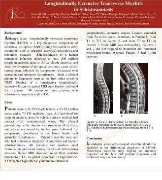
Longitudinally Extensive Transverse Myelitis in Schistosomiasis Juliana M.S.S. Amaral, Igor Ornelas, Natália C. Talim, Lívia E.C.Talim, Rodrigo Kleinpaul, Marcia Prates, Diogo C. Carvalho, Tauana S. Tironi, Izabela Duarte, Anderson Silva, Gisele O Lima, Carolina R. Araujo, Cristiane F. Rocha, Marco A. Lana-Peixoto. CIEM MS Research Center, Federal University of Minas Gerais Medical School, Belo Horizonte, Brazil. a b Background Although acute longitudinally extensive transverse myelitis (LETM) is a key diagnostic component of neuromyelitisoptica (NMO) it may also occur in other conditions such as multiple sclerosis, sarcoidosis and infectious diseases. Schistosomiasis is a human trematode infection affecting at least 200 million people in endemic areas in Africa, South America, and Asia. Involvement of the spinal cord may cause severe lumbar pain followed by paraparesis associated with sensation and sphincter disturbances. Such a clinical picture is frequently seen as the first index event in NMO. Finding of a tumefactive longitudinally extensive lesion on spinal MRI may further confound the diagnosis. We report on three patients with schistosomalmyelitis and LETM. Cases Patients were a 21-YO black female, a 23-YO indian male, and a 70-YO mulattoe male. All had lived for years in endemic areas for schistosomiasis and had had contact with contaminated water. The clinical presentation of the disease was similar in all of them, and was characterized by lumbar pain, followed by paraparesis, dysesthesia in the lower limbs, and sphincter disturbances. Patient 3 was the only one who had a previous diagnosis of intestinal and hepatic schistosomiasis. All patients had positive stool examination and rectal biopsy for ova of Schistosomamansoni. Spinal MRI disclosed in all three patients tumefactive T2- weighted isointense or hyperintense, T1-weighted hypointense gadolinium-enhanced longitudinally extensive lesions. Lesions extended from T4 to the conusmedullaris in Patient 1; from T1 to T12 in Patient 2; and from T7 to T11 in Patient 3. Brain MRI was unrevealing. Patients 1 and 2 did not respond to treatment and remained wheelchair-bound, whereas Patient 3 had a full recovery. Figure. a. Case 2. Tumefactive T2-weighted lesion extending through the thoracic spinal cord. b. Case 3. T2-weighted hyperintense lesion extending from T7 to T11. Conclusion In endemic areas schistosomal myelitis should be included in the differential diagnosis of LETM. Neurologic outcome may depend on immune response of the host but prompt diagnosis and treatment may favor recovery.


