Anatomy of the Neck
1k likes | 2.53k Vues
Anatomy of the Neck. Anterior triangle. Midline of the neck Sternocleidomastoid muscle Lower border of the mandible. Subunits of ant. triangle. Submandibular triangle Submental triangle Carotid triangle Muscular triangle. Submandibular triangle .

Anatomy of the Neck
E N D
Presentation Transcript
Anterior triangle • Midline of the neck • Sternocleidomastoid muscle • Lower border of the mandible
Subunits of ant. triangle • Submandibular triangle • Submental triangle • Carotid triangle • Muscular triangle
Submandibular triangle • Anterior & posterior bellies of digastric muscle • Lower border of the mandible
Submental triangle • Anterior bellies of the digastric muscle • Hyoid bone
Posterior triangle • Anterior border of the trapezius m. • SCM • Middle third of the clavicle
Subunits of post. Triangle • Subclavian triangle • Occipital triangle
Fascial layer of the neck The cervical fascia represents a condensation of connective tissue that extends between anatomic structures
Superficial Fascia • Lies just below the dermis • Deep portions of this layer encase the platysma muscle as well as the voluntary muscles of the face & scalp
Deep cervical fascia • Superficial layer • Middle layer • Deep layer
Superficial layer of deep cervical fascia • Begins from the vertebral spinous processes and splits to enclose the trapezious • Again it splits to invest SCM as well as strap muscle • Superior attachment : occipital protuberance , superior nuchal line & zigomatic arch
Between parotid & submandibular glands the two layer rejoin to form the stylomandibular ligament • Inferiorly the fascia split and attach to the anterior and posterior surface of the sternum : Suprasternal space of Burns
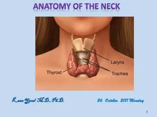
Middel layer of the deep cervical fascia • It encloses the thyroid gland , trachea , pharyngeal constrictor muscle & esophagus • It extends from the hyoid bone down to the sternal attachments and is continuous with fibrous pericardium
Deep layer of the deep cervical Fascia • Anterior to the vertebral bodies • Tips of transverse process • Vertebral spines posteriorly • From the skull base until the coccyx
Prevertebral layer • Alar layer ( until first thoracic vertebra )
Danger space • A potential space is created between the alar and prevertebral fascias because it communicates directly with the mediastinum
Prevertebral space Between the prevertebral fascia and vertebral body
Retropharyngeal space Between alar and the visceral fascia
Tissue space of the neck Between cervical fascia exist potential spaces
Because superficial and deep layers of the deep cervical fascia fuse at the hyoid bone infection in the spaces above the hyoid does not spread directly to spaces below the hyoid
Communication along the entire length of the neck occurs posteriorly along the retropharyngeal and prevertebral spaces .
Submandibular space • Between outer space of of mylohyoid muscle and superficialstructure within submandibular triangle • Along the posterior free edge of the mylohyoid muscle it continuous with the sublingual space • It also communicate with submental and contralateral submandibular space `
Intrapharyngeal space • Inner surface of the superior constrictor muscle and the pharyngeal mucosa • It also known as peritonsillar space
Parapharyngeal space • Medial : superior constrictor m. • Lateral : pterygoid muscles and fascia of the parotid gland • Inferior : Fascial attachment to the hyoid
Posteromedially this space communicates with the retropharyngeal space providing a route to spread infection
Retropharyngeal space • Entire length of the neck • Between visceral fascia and alar fascia • From the skull base down to the T1
Danger space • Between alar fascia and prevertebral fascia • Retropharyngeal space→ danger space →mediastinum
Prevertebral space • Between prevertebral fascial and vertebral column • From the skull base to the lower thoracic area
Common carotid artery • Right side from brachiocephalic artery • Left side from aortic arch • It crosses by omohyoid muscle , , superior & middle thyroid vein
Internal carotid artery • it crosses by hypoglossal nerve , occipital artery & posterior belly of digastric muscle • Near skull base it crosses by glossopharyngeal nerve ,stylohyoid , stylopharyngeous ,styloglossus muscle and styloid process
External carotid artery • It crosses superficially to styloglossus and stylopharyngeous muscle • Terminal branches passing behind the condylar process
Superior thyroid artery • At the level of greater horn hyoid bone • Superior part of thyroid gland , larynx and SCM
Ascending pharyngeal artery • At the level of sup. Thyroid artery posteriorly • Supply pharynx , palate , tonsil , middle ear and meninges
Lingual artery • Above the superior thyroid artery • Runs anterior and superior • Passes beneath the hyoglossus muscle to enter the tongue
Facial artery • On the anterior surface of carotid , deep to the digastric muscle • Passes through the submandibular gland , crosses the inferior border of the mandible • Branches in the neck : ascending palatine artery , tonsillar artery , branches of the submandibular gland , submental artery
Occipital artery • From posterior surface of the external carotid artery the hypoglossal nerve hooks around it • Supply suboccipital region of the scalp , SCM , digastric and stylohyoid muscle
Posterior auricular artery • Posteriorly at the level of the upper border of digastric muscle • Passes between the mastoid and ear • Branches to the parotid gland , auricle and scalp
Terminal branches • Superficial temporal : toward the scalp • Maxillary artery : infratemporal fossa → pterygopalatine fissure → pterygopalatine fossa
Thyrocervical thrunk • Arises from the first part of the subclavian artery just anterior to the scalenus anrerior muscle • Transverse cervical branch → SCM , trapezius • Inferior thyroid artery : deep to the carotid sheath • Supply inferior portion of the thyroid , sup. & inf. Parathyroid gland and a portion of larynx and trachea • Inter the thyroid at the level of cricoid
Internal jugular vein • Sigmoid sinus → intrenal jugular vein → subclavian vein • Major tributaries :inferior petrosal sinus Common facial vein lingual vein superior thyroid vein middle thyroid vein
External jugular vein Posterior auricular vein + posterior branch retromandibular vein Deep to the platysma but superficial to the SCM Terminate in the subclavian vein At its midportion it joined by posterior external jugular vein
Anterior jugular vein • Confluence of the vein in the submandibular region • Drain to the external jugular or subcalavian vein
Glossopharyngeal nerve • Sensory , motor , parasympathic component • It has superior and inferior ganglion • Anterior to the internal and deep to the external carotid artery • Pass between superior and middle constrictor muscle • Innervate tonsil , pharynx and tongue
Tympanic nerve • Arises from inferior ganglion • Tympanic canaliculus → middle ear (jacobson nerve ) • Sensory fiber to the middle ear , eustachian tube and mastoid cavity
Lesser petrosal nerve • Preganglionic parasympathetic fibers • Tympanic plexus → floor of middle cranial fossa → foramen oval → infratemporal fossa → otic ganglion • In the otic ganglion it synapsis with postganglionic fiber of parasympathic that supply the parotid gland
Carotid branch • Arises from IX nerve just below the skull base • Unit with carotid branch of vagus nerve and carries sensory information back from the carotid body and carotid sinus
Pharyngeal branch • Reach to the pharyngeal plexus on the middle constrictor muscle • Sensory innervation


![[PDF] Illustrated Anatomy of the Head and Neck Kindle](https://cdn7.slideserve.com/12506909/slide1-dt.jpg)
![[PDF] DOWNLOAD Illustrated Anatomy of the Head and Neck](https://cdn7.slideserve.com/12519396/slide1-dt.jpg)