Download
1 / 1
10 likes | 273 Vues
Case Report on Neonatal Iron Storage Disease Anushka Kinra, DO, Leanne Mihata, MD, Ali Ghazi-Askar MS, MD Department of Pediatrics, Advocate Hope Children’s Hospital. Etiology:

E N D
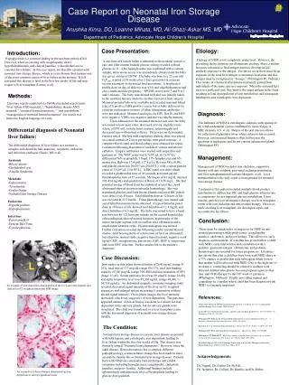
Case Report on Neonatal Iron Storage Disease Anushka Kinra, DO, Leanne Mihata, MD, Ali Ghazi-Askar MS, MD Department of Pediatrics, Advocate Hope Children’s Hospital Etiology: Etiology of NISD is not completely understood. However, the prevailing theory proposes an alloimmune etiology where a mother becomes sensitized to fetal antigen and may develop an IgG antibody response to the antigen. For this to occur there must be an exposure of the fetal liver antigen to maternal circulation and this antigen must be recognized as “foreign.” (Whitington PF, Padmini) This results in a humoral alloimmune (immunity gained from individuals of the same species) response. When the maternal IgG rises to a sufficient titer, they bind to the target antigen in the fetus resulting in fetal dysregulation of iron metabolism and consequent intrahepatic and extrahepatic iron deposition. Case Presentation: Introduction: Hypoglycemia is a common finding in the newborn period of life. However, when co-existing with coagulopathy, direct hyperbilirubinemia, and clinical jaundice, it should alert one to consider liver failure. In this case report, we describe a patient with neonatal iron storage disease, which is a rare disease that remains one of the most common causes of liver failure in the neonate. If left untreated this disease is fatal in the first few weeks of life and may require a liver transplant (Leonis, et al). A one-hour-old female infant is admitted to the neonatal intensive care unit after routine bedside glucose testing revealed a blood glucose of 11. After hypoglycemia was confirmed with a venous sample, intravenous access was immediately obtained and the baby was given a bolus of D10W. The baby was born to a 32 year old G2P2001 woman at 38 weeks plus 2 days gestation via cesarean section secondary to decreased fetal movement. A biophysical profile done on day of delivery was 4/10, and oligohydramnios had also complicated the pregnancy. APGAR scores were 7 and 9 at 1 and 5 minutes. The baby transitioned well and was initially taken to the newborn nursery while the mother recovered from surgery. Maternal prenatal labs were available and revealed maternal blood type of A positive, GBS positive screen, but as baby delivered via cesarean section prior to onset of labor, intrapartum antibiotics were not indicated. Maternal serologies for HIV, Hep B, and HSV were negative; VDRL was negative and she was rubella immune. Upon admission to the neonatal intensive care unit, the baby was noted to have poor color, decreased activity, slightly icteric sclera, a II/VI soft, systolic heart murmur, splenomegaly and decreased tone with normal reflexes. There were no dysmorphic features noted. She had mild respiratory distress, thus, was placed on a nasal cannula at 2 liters per minute with good response. A complete blood count and blood culture were obtained for sepsis evaluation following placement of umbilical venous and arterial catheters. Empiric antibiotics were started with ampicillin and gentamicin. The WBC count was 6.7x10³/µL (6.7x10⁹/L), differential 76% neutrophils, 1 band, 11% lymphocytes and 6% monocytes, Hgb was 17.4 g/dL (17.4 g/L), Hct was 48% (0.48), and platelet count was 29x103/µL (29x109/L), with a repeat platelet count of 15x103/µL (15x109/L). A DIC panel was done which revealed a prothrombin time of 36 seconds, activated partial thromboplastin time of 57 seconds, fibrinogen of 43 mg/dL (normal 203-424 mg/dL) and quantitative d-Dimer of 10.58 mg/L. She had minimal oozing of blood from her umbilical arterial line; head ultrasound showed no intraventricular hemorrhage. She was transfused platelets and fresh frozen plasma and serial DIC panels were done every 8 hours. Total bilirubin done at 12 hours of life was elevated at 12.2 mg/dL. Triple phototherapy was started and serial bilirubin measurements obtained. A repeat bilirubin panel done at 18 hours of life showed total bilirubin of 12.4 mg/dL and direct bilirubin of 6.5 mg/dL. The baby was noted to have a heart rate between 86-121 beats per minute on the second hospital day; echocardiogram showed normal function, hypertrophy of the intraventricular septum with no outflow tract obstruction and a small patent foramen ovale. Electrocardiogram was normal. Further evaluation revealed the following results: normal thyroid studies; mild heterogeneity of echotexture of liver on ultrasound, but otherwise normal study; negative hepatitis panel, negative work up for CMV, toxoplasmosis, parvovirus, EBV, HHV 6, enterovirus and acute HSV infection. Further studies led to the patient’s diagnosis. Methods: Literature search conducted via PubMed included search terms “liver failure AND neonatal,” “hepatobiliary disease AND neonatal,” “neonatal hemochromatosis,” “iron metabolism,” “management of neonatal hemochromatosis” our search was limited to English language text only. Diagnosis: The hallmark of NISD is extrahepatic siderosis with sparing of the retiuloendothelial system confirmed by tissue biopsy or MRI. (Knisely A.S. et al). Biopsy of the oral mucosa allows for collection of glandular tissue where siderosis has occurred. However, oral mucosal biopsies often fail because the specimen is inadequate and do not contain submucosal glands (Whitington PF). Management: Management of NISD includes iron chelation, supportive therapy with anti-oxidants, post-natal exchange transfusion, and liver transplantation (Escolano-Margarit, et al). Liver transplantation is the only curative management for neonatal iron storage disease. Treatment of this patient included multiple blood product transfusions to address her DIC and high glucose infusion rate to compensate for her impaired glucose metabolism. Upon transfer, patient received intensive therapy at a liver transplant center with iron chelation and anti-oxidant therapy. However, while awaiting liver transplant, she developed sepsis and succumbed to her illness. Differential diagnosis of Neonatal liver failure: The differential diagnosis of liver failure in a neonate is complex and should include anatomic, metabolic, endocrine and infectious etiologies (Saenz, MS et al). Anatomic -Biliary atresia -Choledochol cyst -Alagille Syndrome Metabolic -Galactosemia -Tyrosinemia -Crigler-Najjar -Neonatal Iron Storage Disease Endocrine -Hypothryoidism -Hypopituitarism Infectious -Parvovirus B19 -Epstein-Barr Virus -Cytomegalovirus Conclusion: There must be a high index of suspicion for NISD in any neonate presenting with hypoglycemia, coagulopathy, jaundice, cholestasis, and poor feeding. This allows for early diagnosis and treatment. If a mother has given birth to a child with NISD, early intervention and consultation with a pediatric gastroenterologist, obstetrician, and pediatric hematologist are essential for future pregnancies. Literature has shown that after a child has been born with NISD, there is a 75% chance or greater that each subsequent infant born to that mother will be affected with NISD. Due to the high rate of recurrence, counseling should be offered to mothers with affected children who plan to become pregnant again so they may start IVIG therapy by the 18th week of gestation (Whitington, Hibbard). Finally, providing support and counseling for a mother whose child has been diagnosed with NISD is extremely important. References: Leonis MA, Balistreri WF. Neonatal hemochromatosis: it’s OK to say “no” to antioxidant-chelator therapy. Liver Tranpl 2005;6:880-4 Saenz MS et al. Neonatal liver failure: a genetic and metabolic perspective. Current Opinion in Pediatrics 2010, 22: 241-245 Whitington PF, Padmini M. Neonatal Hemochromatosis: Is it an alloimmune disease? J Pediatric;Gastroenterology and Nutrition 2005; 40: 544-9 Knisely A.S. et al. Neonatal hemochromatosis. Gastroenterol Clin N Am 32 (2003) 877-889 Whitington PF. Fetal and infantile hemochromatosis. Hepatology 2006; 43:654-660 Whitington, PF, Hibbard JU. High-dose immunoglobulin during pregnancy for recurrent neonatal hemochromatosis. The Lancet. Vol 364 Iss 9446 1690-1698 Escolano-Margarit, MV et al. Exchange Transfusion as a Possible Therapy for Neonatal Hemochromatosis. JPGN. Vol 50 Num 5 May 2010 Case Discussion: Iron studies in this infant showed ferritin of 2643 ng/mL (range 8-252), total iron of 177 mcg/dL (range 50-177), total iron binding capacity of 186 mcg/dL (range 100-400) and iron saturation of 95% (range 15-45). Serum ammonia level was 89 µmol/L (range 28-88), and alpha fetoprotein level was 87,328 ng/mL (range 48,406 +/- 34,718 ng/mL). An abdominal magnetic resonance imaging study revealed decreased signal intensity of the liver on T2-weighted sequences and enlarged spleen measuring 8 centimeters without altered signal intensity. On in-phase images, the liver intensity decreased, which was suggestive of iron deposition. The pancreas appeared normal. A buccal biopsy was done to evaluate for iron deposition in the salivary glands, but no salivary glands were visualized. The child was transferred to a liver transplant center with the presumed diagnosis of neonatal iron storage disease (NISD). An example of iron deposition demonstrated in the liver and extra-hepatic sites (spleen) on T2 weighted abdominal MRI image TheCondition: Neonatal iron storage disease is a severe liver disease associated with both hepatic and extrahepatic iron deposition, leading to liver failure within the first few weeks of life. This disease was formerly termed “Neonatal hemochomatosis.” However, since the adult disease, Hemochromatosis has a complete different pathophysiology, a nomenclature change has been made to more accurately classify this as Neonatal iron storage disease. Patients born with NISD are commonly born premature and exhibit symptoms including hypoglycemia, coagulopathy, cholestatic jaundice, and poor feeding. Additional findings include splenomegaly and pancreatic islet cell hyperplasia leading to glucose dysregulation. Acknowledgements: Dr. Nagpal, Dr. Alattar, Dr. McFall, Dr. Jazmines, Dr. Collins, Dr. Ramilo, and Dr. Mehta An example of a buccal biopsy demonstrating iron deposition in salivary glandular tissue