Overview of Hearing and Its Physiology
Learn about the anatomy, physiology, and testing methods of hearing. Explore the auditory pathway and when to suspect hearing problems.

Overview of Hearing and Its Physiology
E N D
Presentation Transcript
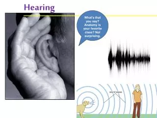
Introduction Hearing is the ability to perceive sound by detecting vibrations through an ear. It is also called Audition It is a type of Mechanoreceptor sense
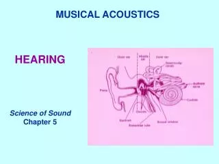
Anatomy of EAR The ear is formed of three anatomical and functional parts: • Outer ear – functions in hearing • Middle ear – functions in hearing • Inner ear – functions in both hearing and equilibrium
Outer (external) Ear • Involved in hearing only • Structures of the external ear • Pinna (auricle) • External auditory canal • Tympanic membrane
The Middle Ear or Tympanic Cavity • Air-filled cavity within the temporal bone • Only involved in the sense of hearing • 3 bones (ossicles) : Malleus , Incus & Stapes • 2 muscles Tensor tympani & Stapedius • The Eustachian tube
Inner Ear or Bony Labyrinth • Includes sense organs for hearing & balance • Filled with perilymph • Bony labyrinth cochlea vestibule 3 semicircular canals • Membranous labyrinth
Organ of hearing Organ of Corti • The receptor epithelium for hearing • Located within the cochlea • Hearing receptors hair cells on the basilar membrane • Gel-like tectorial membrane is capable of bending hair cells • Cochlear nerve attached to hair cells transmits nerve impulses to auditory cortex on temporal lobe
Physiology of Hearing The auricle directs sound waves into the external auditory canal. Sound waves strike the tympanic membrane causes it to vibrate back and forth. The distance it moves depends upon the intensity and frequency of the waves. The central eardrum connects to the Malleus, which also starts to vibrate. This vibration is then transmitted to the Incus and Stapes. As the stapes moves back and forth, it pushes the membrane of the oval window in and out.
Cont… The movement of the oval window sets up fluid pressure waves in the perilymph of the cochlea. Pressure waves are transmitted to the round window, causing it to bulge outward. These waves in turn create pressure waves in the endolymph of the cochlear duct. This causes the basilar membrane to vibrate, which moves the hair cells leading to receptor potentials and ultimately nerve impulses.
Auditory Pathway Sound waves are transmitted as electrical waves through the 8th nerve to the cochlear nuclei on each side of medulla (which has two nuclei dorsal and ventral cochlear nucleus) Fibers from ventral nucleus (which is concerned in time difference) will relay into the superior olivary complex in the pons , and some little fibers from dorsal cochlear nucleus will relay there too.
Auditory Pathway Most fibers from the dorsal cochlear nucleus (concerned in quality of sound) go directly to the inferior colliculus of midbrain. The inferior colliculus also receives fibers from superior olivary nucleus. The lateral lemniscus is a tract of axons in the brainstem that connects the previous nuclei together and carrying auditory signal in-between.
Auditory Pathway From the inferior colliculus Then fibers go to the medial geniculate body in the thalamus, then to the primary auditory cortex in the temporal lobe. The area in cortex concerned with hearing situated in the superior temporal gyrus (Brodmann’s area 41,42).
When hearing tests are required? Hearing tests are performed in the conditions mentioned below. Hearing impairment As a part of routine assessment of child development Maybe a part of general medical examination Maybe used to know the cause of Tinnitus
When to suspect hearing problems? • Do you have a problem hearing over the telephone? • Do you hear better through one ear than the other? • Do you have trouble following the conversation with two or more people talking at the same time? • Do people complain that you turn the TV volume up too high? • Do you have dizziness, pain, or ringing in your ears? • Do you have trouble understanding the speech of women and children?
Hearing impairment Hearing impairment : Diminution in the acuity of hearing Types of hearing loss: Conductive hearing loss. Perceptive or sensorineural hearing loss. Mixed type.
How hearing tests are done? Hearing of a person can be tested by two methods: • Clinical tests • Audiometric tests
A . Clinical Tests • Finger Friction Test • Watch Test • Speech Test • Tuning Fork Tests
Finger Friction Test Is a quick and rough method of testing hearing by rubbing the thumb and the finger close to the patients ear and ask if the sound is listened.
Watch Test Is not done now but had been very popular before the invent of audiometers. It's done by bringing a clicking watch close to the ear and measuring distance at which it is heard
Speech Test The patient stands at a distance of 6 meters from the examiner. The examiner says words and gradually walks towards the patient. The distance at which conversational & whispered voice are heard is measured. For clinical purposes 6m is considered normal for both conversational and whispered speech. While performing the test eyes of the patient are covered to prevent lip reading, also the other ear is blocked.
Tuning Fork Tests Theses tests are performed with the tuning forks of different frequencies such as 128,256,512 Hz For routine practice, tuning fork of 512 Hz is ideal
Tuning Fork Tests • The clinically useful tuning forks tests include: • Rinne Test • Weber Test • Others
Rinne Test In this test air conduction AC of the ear is compared with its bone conduction BC A vibrating tuning fork is places on the pt mastoid and when he stops hearing it brought beside the meatus. If he still hears, AC is more than BC
Interpretations Rinne test is called positive when AC is longer and than BC. It is seen in normal person or those having sensorineural deafness. A negative Rinne test when BC > AC . Is seen in conductive deafness
Weber Test In this test , a vibrating tuning fork is placed in the middle of the forehead and the pt is asked in which ear the sound is heard. Normally it is heard equally in both ear
Interpretations It is lateralized to the worse ear in conductive deafness and to the better ear in sensorineural deafness
B. Audiometric tests Audiometry is the measurement of the sense of hearing. An audiometer is an electronic machine that produces pure tones, the intensity of which can be increased or decreased in 5db steps. A graph or audiogram is developed by this way that shows the hearing threshold
Examination of the external ear by Otoscope An Otoscope is a medical device which is used to look into the ears. Health care providers use otoscopes to screen for illness during regular check-ups and also to investigate when a symptom involves the ears An Otoscope consists of three parts: The handle which contains the power for the light source. The head which contains the light bulb and magnifying lens. The cone which is inserted into the ear canal
Using the Otoscope The examiner first straightens the ear canal by pulling on the pinna , inserts the ear speculum into the external ear. The examiner can then look through a lens on the rear of the instrument and see inside the ear canal. Diseases which may be diagnosed by an otoscope include otitis media and otitis externa, infection of the middle and outer parts of the ear.