Lymphocytic Gastritis
330 likes | 929 Vues
Once you have mastered H pylori gastritis and the atrophic gastritides , there are only a few more gastritides about which we know anything. Lymphocytic Gastritis. At low power, there is inflammation, mainly superficial. Check out the mess in the surface epithelium!.

Lymphocytic Gastritis
E N D
Presentation Transcript
Once you have mastered H pylori gastritis and the atrophic gastritides, there are only a few more gastritides about which we know anything
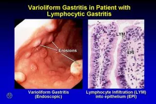
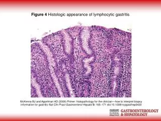
At low power, there is inflammation, mainly superficial. Check out the mess in the surface epithelium!
Some cases of lymphocytic gastritis have the giant folds and protein losing of Menetrier’s disease
Small patches of lymphocytic gastritis like this pop up next to all kinds of things
Lymphocytic Gastritis • Clinical Aspects • Endoscopic: • nothing, polyps, giant folds • Clinical: • nothing, sprue-associated, • Menetrier’s syndrome, • H pylori elsewhere
FOCAL …ITIS • Very, very common • More common after search • More common if there are residents • The entire gut is at risk • Almost never diagnostic • Annoying • The more foci the better!
Often mainly lymphocytes with PMNs, macrophages and epithelial damage
Was found often in patients with Crohn’s Then it was found in patients with UC
Every so often a granuloma or 2 may be part of focal gastritis
At low power, the dominant change is the prominence of the PITS
The high N:C resembles dysplasia PITS, PITS and more PITS, often serrated contours
This change is an expansion of the proliferative zone (actually, the neck region, not the pits)in compensation for surface epithelial injury. This is very common in our practice. Most of the time, we do not know the cause.
Stomach Small bowel Originally described on the gastric side of a gastro-enteric anastomosis, due to bile reflux
In almost 20 year old studies of patients with chronic NSAIDs use. Not much change recently. Gastric erosions occur in 40-60% Gastric ulcers occur in 10-30% Duodenal ulcers occur in 5%