Download
1 / 29
290 likes | 565 Vues
Psychology 4051. The Retina and LGN. Retino-Geniculate-Cortical Pathway. The Retina. Located at the back of the posterior chamber, forms the inner tunic of the eye. Surface on which the visual image is focused. The Retina. A laminar tissue with multiple layers.

E N D
Psychology 4051 The Retina and LGN
The Retina • Located at the back of the posterior chamber, forms the inner tunic of the eye. • Surface on which the visual image is focused
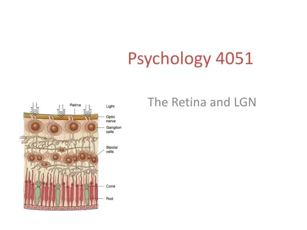
The Retina • A laminar tissue with multiple layers. • Pigment epithelium, photoreceptor layer, external limiting membrane, outer nuclear layer, outer plexiform layer, inner nuclear layer, inner plexiform layer, ganglion cell layer, optic nerve layer, internal limiting membrane. • Transduction takes place in the photoreceptors. • Rods and cones
Light hits the outer segment of rods and cones which contain photosensitive chemicals (photopigments). • Rod-rhodopsin cones-iodopsin • The light changes the molecular properties of the photopigments, which in turn changes the electrical state of these cells – this is called transduction.
The Retina The design of the retina is unusual. Light must pass through 8 layers Before it hits the Photoreceptors. Light
The Retina • The retina contains a central pit, above which cell layers are pushed away. • The fovea: 1mm in diameter • Packed with cones
fovea • Fovea • At the center of the retina, high acuity • Reduced light distortion
The Retina • Rods and cones connect with horizontal and bipolar cells (collector cells). • Lateral interaction takes place at horizontal cells. • Bipolar cells are connected to retinal ganglion cells. • Amacrine cells connect adjacent bipolar cells.
Rods • 120 million rods in the human retina • Concentrated in the periphery of the retina
Rods • Extremely sensitive to light. • Many rods converge onto a single retinal ganglion cell (RGC)
Rods – good light sensitivity • As a result, low levels of light can be detected • Responsible for night vision • But, spatial resolution is very poor • RGCs can not determine which rods were stimulated
Cones • Approximately 6 million cones • Cones are packed densely in the fovea
Cones • Cones are connected to bipolar cells, then to retinal ganglion cells (RGCs) • Few cones converge onto a single retinal ganglion cell (RGC) • As a result, resolution is excellent
Cones • Light sensitivity is poor • Day vision
Cones • Humans have 3 different cone types, each with a different photopigment. • Photopigments are maximally sensitive to specific wavelengths. • Short wavelength sensitive • Mid-wavelength sensitive • Long wavelength sensitive
Retinal Ganglion Cells • Retinal ganglion cells possess receptive fields that are responsive to light stimulation. • Receptive field – that area of the retina over which a ganglion cell is sensitive to light stimulation (area of the retina that the RGC monitors).
Receptive Fields • Their receptive fieldsare designed in an antagonistic fashion • A single receptive field has an on-center/off- surround arrangement or an off-center/on-surround arrangement.
“On-Center” Cells • On-center cell: Light stimulation of the center of thereceptive field produces depolarization and an increase in the firing rate of the ganglion cell. • Stimulation of the surround produces a hyperpolarization and a decrease in the firing rate of the cell. - +
“Off-Center” Cells • Off-center cell: Light stimulation of the surround of thereceptive field produces depolarization and an increase in the firing rate of the ganglion cell. • Stimulation of the center produces a hyperpolarization and a decrease in the firing rate of the cell. - +
Retinal Ganglion Cells • The axons of the RGCs converge and leave the eye through as the optic nerve. • They connect to the lateral geniculate nucleus (LGN).
Retinal Ganglion Cells • Most RGCs fall into two functional classes, M and P cells • M cells – project to the magnocellular layers of the lateral geniculate nucleus (LGN) • P cells – project to the parvocellular layers of the LGN
M Cells • M cells receive input from a relatively large number of photoreceptors (mostly input from rods) • Good light sensitivity, good temporal resolution (sensitive to motion) • Poor spatial resolution • Large – with broad axons and consequently faster nerve conduction velocities • Not color-sensitive
P Cells • P cells receive input from a relatively small number of photoreceptors (mostly input from cones) • Good spatial resolution • Poor temporal resolution • Colour sensitivity
LGN • A six-layered thalamic relay station
LGN • Cells in the magnocellular layer have large cell bodies and long, straight axons. • Cells in the parvocellular layer possess smaller cell bodies and short, curved axons. • Layers 1, 4, and 6 receive input from the contralateral eye. • Layers 2, 3, and 5 receive input from the ipsilateral eye.
LGN • Most parvocellular cells appear to be responsive to color and have center/surround receptive fields.