Download
1 / 12
120 likes | 297 Vues
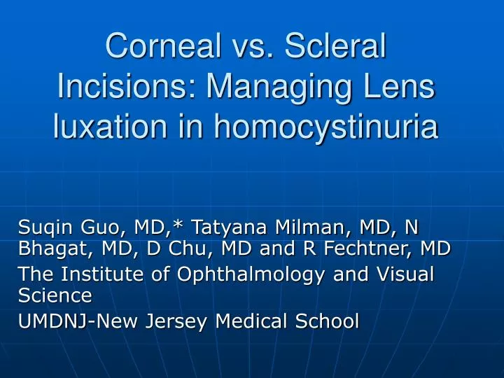
Corneal vs. Scleral Incisions: Managing Lens luxation in homocystinuria. Suqin Guo, MD,* Tatyana Milman, MD, N Bhagat, MD, D Chu, MD and R Fechtner, MD The Institute of Ophthalmology and Visual Science UMDNJ-New Jersey Medical School. Corresponding /Senior Author Suqin Guo, MD.

E N D
Corneal vs. Scleral Incisions: Managing Lens luxation in homocystinuria Suqin Guo, MD,* Tatyana Milman, MD, N Bhagat, MD, D Chu, MD and R Fechtner, MD The Institute of Ophthalmology and Visual Science UMDNJ-New Jersey Medical School
Corresponding /Senior AuthorSuqin Guo, MD • Assistant professor • The Institute of Ophthalmology and Visual Science • UMDNJ-New Jersey Medical School
Purpose of the Study • To compare the wound healing of clear corneal incision with scleral incision in surgically managing lens luxation in a child with homocystinuria
Methods-Case Report • A 10-year old child with a known history of homocystinuria • Presented with bilateral complete luxation of the lens into the anterior chamber • Recurrent angle-closure glaucoma from pupillary block of complete luxated lens into anterior chamber.
Case Report • Her Intraocular pressure was medically uncontrollable. • Her left eye: Underwent lensectomy and anterior vitrectomy via clear corneal incision by an anterior segment surgeon • Her right eye: Had pars plana lensectomy ( PPL) and vitrectomy (PPV) by a vitreoretinal specialist
Results-surgical OutcomeLEFT eye- lens removal via clear cornea • Her left eye underwent Lensectomy via a clear corneal incision and healed well • Intraocular pressure remained within normal limit without needing any medication over 7 years. • 20/80 with aphakic correction
Results-surgical OutcomeRIGHT eye – post-PPV+PPL • Her right eye that underwent pars plana lensectomy (PPL) and vitrectomy (PPV) via sclerotomy incisions • Developed scleral necrosis, scleromalacia over the sclerotomy sites • Had poor controlled intraocular pressure (IOP)
Results-surgical OutcomeRIGHT eye – post-PPV+PPL • Her right eye needed multiple scleral patch graft surgeries
Results-surgical OutcomeRIGHT eye – post-PPV+PPL • Her right eye, later, perforated over the sclerotomy sites. Additional multiple operations were needed, including scleral patch graft and retinal detachment repair surgeries. • Visual acuity= HM
Discussion/Conclusion • One of the main characteristics of homocystinuria is a high risk of arterial and venous thromboembolism. • High risk of tissue necrosis secondary from ischemia • High risk of general anesthesia
Conclusion • Sclera is vascular tissue whereas cornea is avascular. Arterial and venous thromboembolism could occur within scleral vessels due to minimal surgical trauma or ischemic changes from abnormal IOP, causing poor wound healing.
Discussion/Conclusion • Cornea is avascular and may be spared from thromboembolism. Clear corneal incision may provide better wound healing in patients with homocystinuria.