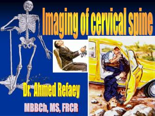
Dr. Ahmed Refaey
Imaging of cervical spine. Dr. Ahmed Refaey. MBBCh, MS, FRCR. 3% of MVA patients , have cervical spine injury 10-20% patients with head injury, have also cervical spine injury Most cervical spine fractures occur at two levels

Dr. Ahmed Refaey
E N D
Presentation Transcript
Imaging of cervical spine Dr. Ahmed Refaey MBBCh, MS, FRCR
3% of MVA patients , have cervical spine injury • 10-20% patients with head injury, have also cervical spine injury • Most cervical spine fractures occur at two levels • 17% of patients have a missed or delayed diagnosis with a risk of perminant neurologic damage • 1/3 of injuries occur at level of C2 and ½ occur at level of C6-C7
The NEXUS criteria state that a patient with suspected cervical spine injury can be cleared providing the following: *- no posterior midline cervical spine tenderness *- no evidence of intoxication is present *- patient has a normal level of alertness *- no focal neurologic deficit
Plain films • 3 views should be taken : - true lateral view ( must include all 7 cervical vertebrae as well as the C 7 – T1 junction ) - an AP view - an open mouth (odontoid view)
The lateral view … • The lateral view is the most useful view, approximately 85-90 % of spinal injuries are evident on this view • Should be obtained & examed before any other films are taken • All 7 cervical vertebrae and the C7-T1 junction must be visualized because the cervicothoracic junction is a common place for traumatic injury
AP & odontoid view • If lateral view is normal, proceed with AP & odontoid views • Patient should be maintained in cervical immobilization and plain films or CT scans obtained untill all vertebrae are clearly visible.
Systematic approach: * check alignment by following 3 contour lines - anterior contour line - posterior contour line - spinolaminar contour line
These lines should follow a slightly lordotic curve, smooth and without step-offs. • Any malalignment should be considered evidence of ligamentous injury or occult fracture and cervical spine immobilization should be maintained untill a definitive diagnosis is made.
AP view • Alignment should be evaluated using the edges of vertebral bodies and articular pillars • Hight of vertebral bodies should be equal • Hight of joint space should be equal • Spinous process should be in midline, if displaced to one side , a facet dislocation should be suspected.
Odontoid view • The distance from the dens to lateral masses of C1 should be equal bilaterally. • Any asymmetry is suggestive of a fracture of C1 or C2 • Lateral mass of C1 should line up with lateral margins of supriorarticular facet of C2. if not, a fracture of C1 is suspected.
Prevertebral space • Nasopharyngeal space {C1} -- 10 mm in adult • Retropharyngeal space {C2-C4} -- 5-7 mm • Retrotracheal space {C5-C7} -- 14mm in children -- 22 mm in adults
Prevertebral soft tissue swelling is important as it is usually due to hematoma 2ry to occult fractures • Soft tissue swelling in symptomatic patient should be considered an indication for further radiographic evaluation
CT • 20% of fractures are missed on plain radiographs • Useful in fractures that result in neurologic deficit and in fractures of posterior elements ( e.g. Jefferson’s fracture)
Advantages of CT • Excellent in identifying osseous compromise of the vertebral canal • Visualization of subtle fractures • Provides patient comfort by being able to reconstruct images in axial , sagittal , coronal planes and 3D from one patient position. • Soft tissue window & bone window
****** limitations: * unable to show ligamentous injuries * relatively high costs
Excellent in identifying osseous compromise of the vertebral canal
Provides patient comfort by being able to reconstruct images in axial , sagittal and coronal planes from one patient position as well as 3-d reconstruction
MRI • Advantages: * excellent soft tissue contrast, making it the study of choice for spinal cord survey , hematoma and ligamentous injuries. * good general overview because of its ability to show informations in different planes * ability to demonstrate vertebral arteries which is useful in evaluating fractures involving the course of vertebral arteries * no ionizing radiation
MRI • Disadvantages * loss of bony details * high cost - patients with pacemakers and certain ferromagnetic materials ( aneurysm clips ) are not able to be scanned
MRI diagnostic values • Spinal cord lesions • Bone marrow pathology • Ligamentous injuries • soft tissue edema
MRI • T1W – display anatomic details • T2W – display pathologic changes better • Together – enable detection & characterization of most lesions
Spinal stability is depending on at least two intact columns. • When two of the three columns are disrupted , it will allow abnormal segmental motion.
Spinal cord injury • Two types: * non-hemorrhagic: only high signal on T2W * hemorrhagic: areas of low signals on T2W within the area of edema
Hemorrhagic spinal cord injury has an extremely poor outcome
Hyperflexion Hyperextension Compression
Hyperflexion ……. • Excessive flexion of neck in sagittal plane • Diving in shallow water • Flexion tear drop fracture