Download
1 / 16
430 likes | 2.05k Vues
Embryology Development of Dermatomusculoskeletal System. Biology Medic Department 2011. SKELETAL MUSCLE DEVELOPMENT. The Muscular system mesoderm (exc. iris musc.neuroectoderm , esop.musc . smooth musc .) Paraxial Mesoderm somites

E N D
Embryology Development of Dermatomusculoskeletal System Biology Medic Department 2011
SKELETAL MUSCLE DEVELOPMENT • The Muscular system mesoderm (exc. iris musc.neuroectoderm, esop.musc. smooth musc.) • Paraxial Mesoderm somites A. Ventromedial (Sclerotome) Vertebrae+ribs B. Dorsolateral (Dermomyotome) myotome (myoblast) + dermatome (dermis-fibroblast) • Somatic Mesoderm +Ventral dermomyotome of somites +Neural crest cells Mesenchymemyogenesis Induction by MyoD gene elongation of the nuclei& cell bodies Myoblast myotubes+ myofilament+ myofibril invested with ext.lamina.endomysium • Fibroblastperimysium and epimysium • Dev before birth, sizediameter fiber, length&wide to grow with skeleton
Myotomes 1. Epaxial the extensor muscles of the neck & vertebral column, Caudal myotom regressed 2. Hypaxial cervical myotomes scalene, pravertebral, geniohyoid & infrahyoid m. • thoracic myotomes lateral and ventral flexor muscles of the vertebral column • lumbar myotomes quadratuslumborum m. • Pharyngeal arch m. facial expression,m. of mastication, pharynx and larynx • Mesenchymal cells near the prechordal plate Ocular musclesextrinsic eye muscles • Myoblast surrounding the developing boneslimb muscles • Occipital myotomTongue Muscles
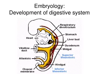
SKIN DEVELOPMENT • EctodermEpidermis • MesodermmesenchymeConnective tissues in the dermis • Epidermis • 1st and 2nd trimesters of pregnancythickness • PrimordiaEctodermal cells at 4 weeks Proliferation (Periderm) & Basal/germinativum layer • Basal layerStr, germinativumproduced more cell to superficial layerIntermediate layer (11 weeks) Epidermal ridgesgrooves(10-17 weeks) • Peridermkeratinization,desquamation(Vernixcaseosa)--<Str, Corneum (21 weeks) • Ektoderm Neural crest cellmigrate into the mesenchyme of the developing dermis Melanoblastmigrate to dermoepdermaljunctionMelanocyte
Glands of The Skin Development 1. Sebaceous Gland • Derived from the epidermis to dermis • ( develop from epithelial root sheath of hair follicleprimordiacentral alveoli ruptureoil excretion)
Glands of The Skin Development 2. Sweat Glands • Epidermal down growth to the mesenchyme • Cellular bud of sweat glandelongate Central cell degenerate lumentheperipheral cells secretory & Myoepithelial cells
Hair Follicles Development • Proliferation of str. Germinativum to the dermishairbudshair bulb(primordia) epithelial cells(germinal matrix)produces hair • Hair bulbs invaginated by small mesenchyme hair pappillae • The peripheral cellsepithelial root sht. • Surrounding messenchymal diff. cellsdermal root sht. Arrector m. attach to the dermal root
Skeletal System Development • Sclerotomevertebrae & ribs. • Mesodermalcellsmesenchym(Embryonic con. tissue)condensation (gene act.)osteogenesis • Mesenchymepre existing membranous sheaths Intramembranous bone formation • MesenchymalcartilagesEndochondral bone formation • Signaling mol.BMP-5,BMP-7
Histogenesis of cartilage • 5thweekmesenchymecondensechondrification centres diff. chondroblast collagenousfibril&ground substance(ECM). Histogenesis of Bone • Intramembranous Ossification mesenchymecondenseshighly vascular some cells diff.osteoblast & deposit unmineralized) osteon matrix (osteoid) osteocytesSpicules of bone concentric lamella(layers) • EndochondralOssificationcartilagelong bone(primary centres-diaphysis) chondrocytes hypertrophythe matrix calcified centre cells die periosteum invasion by vasc. conn,.Tissuesome cells diffhemopoetic cells. • Lengthening growth platesSec. oss.in the epiphysisfew years after birth • Ossification of limb bonesat the end of the embryonic perioddemands supply Ca&P
Development of Vertebral Column • Mesenchymal cellssclerotomes3 main areas: around notochord, neural tube and body wall. • Frontal sectpaired condensation of mesenchymal areas around notochordloosely arr. Cranially& densely packed cells caudally. • Some densely packed cellsmovecraniallyooposite the centres of myotomesIv disc • The remaining cells fusecentrumbody of vertebrae( each centrum dev from 2 sclerotomesintersegmentalstructuresinter. A. lie in each side vert. bodies.
Development of Vertebral Column… • Notochord degeneratesgelatinouscentrenucleuspulposus • Surroundedcircularly arranged fiberannulusfibr. • Messenchymal cells surrounding neural tubeneural a • 6thweekchondrificationcentresappearneuralarch+centrumfusespinous&transverseprocextensionoss. Vertebrae begins in the embryonicend by the 25th year
Ribs Development • Mesenchymal costal procoriginal site of union costal pros with vertreplacedcostovertebral synovial joints Sternum Development • A pair of vertical mesenchymal bands(sternal bars)develop ventrolaterallymovemediallychondrificationfusecraniocaudallyformmanubrium, sternebrae&xiphoid proc(oss.childhood) • .
Development of Vertebral Column • Mesenchymal cellssclerotomes3 main areas: around notochord, neural tube and body wall. • Frontal sectpaired condensation of mesenchymal areas around notochordloosely arr. Cranially& densely packed cells caudally. • Some densely packed cellsmovecraniallyooposite the centres of myotomesIv disc • The remaining cells fusecentrumbody of vertebrae( each centrum dev from 2 sclerotomesintersegmentalstructuresinter. A. lie in each side vert. bodies.
Development of the Cranium • Develop from mesenchymearround the developing brain, consist of: - The neurocraniumprotective case for the brain Cartilaginous base of craniumfusionendochondralosssequence:occ.bone, sphenoid, ethmoid Membranous neurocranium: Intramembranousoss. messenchymearround & top of the braincalvariafontanelles(suture meet)
The viscerocraniumskeleton of the face • Cartilaginous: neural crest cellsmigrtaes from pharyngeal arches.Hox 1stmalleus incus, • 2nd stapes+temporalbones,lesserhorn,sup sphenoid bone, • 3rd 4th,6th greater horns+inf part hyoid bone • .4thlaryngeal cartilages • Membranous:max. prominencesquamous, temporal,zygomatic and maxillary, • Mandibularprominencemandible
. Terimakasih