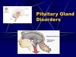
Pituitary Gland Disorders
270 likes | 881 Vues
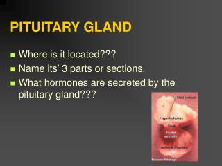
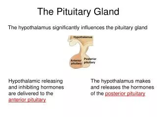
Pituitary Gland Disorders. Diabetes Insipidus (DI) is very different from the disease called sugar diabetes (diabetes mellitus)

Pituitary Gland Disorders
E N D
Presentation Transcript
Pituitary Gland Disorders • Diabetes Insipidus (DI) is very different from the disease called sugar diabetes (diabetes mellitus) • DI is caused by the insufficient release of ADH from the neurohypophysis. Without ADH acting on the collecting ducts in the kidneys, the normal urine output of 1–1.5 liters per day increases to over 2.5 liters per day and dehydration and hypernatremia results
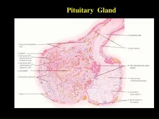
The Thyroid Gland The butterfly-shaped thyroid gland is located inferior to the larynx and anterior to the trachea. It has two laterally placed lobes separated by a bridge-like isthmus
The Thyroid Gland • Most of the thyroid gland is composed of spherical groups of follicular cells called thyroid follicles • The follicles store a 100-day supply of its two hormones in an inactive gel-like substance called TGB (for thyroglobulin)
Thyroid Hormones • TGB is a large glycoprotein made from the oxidation and iodination of molecules of the amino acid. tyrosine • The two hormones released from TGB are: • thyroxine or T4 (tetraiodothyronine) • and T3 (triiodothyronine)
Thyroid Hormones • In the blood, T3 and T4 are bound to pre-albumins, albumin, and a specific carrier protein called thyroid-binding globulin (TBG) • Most T4 released from the thyroid is converted “peripherally” (by enzymes in the blood) into T3 , which is a more active hormone • Together with hGH and insulin, thyroid hormones accelerate body growth, particularly the growth of the nervous and skeletal systems
Thyroid Hormones • Thyroid-stimulating hormone (TSH) is released by the anterior pituitary gland in response to TRH secreted into the portal system • The hypothalamus responds to higher circulating levels of T3 and T4 via negative feedback to inhibit TRH secretion
1 1 1 1 1 Low blood levels of T3 and T3 or low metabolic rate stimulate release of Low blood levels of T3 and T3 or low metabolic rate stimulate release of Low blood levels of T3 and T3 or low metabolic rate stimulate release of Low blood levels of T3 and T3 or low metabolic rate stimulate release of Low blood levels of T3 and T3 or low metabolic rate stimulate release of Hypothalamus Hypothalamus Hypothalamus Hypothalamus Hypothalamus TRH TRH TRH TRH TRH 2 2 2 2 TRH, carried by hypophyseal portal veins to anterior pituitary, stimulates release of TSH by thyrotrophs TRH, carried by hypophyseal portal veins to anterior pituitary, stimulates release of TSH by thyrotrophs TRH, carried by hypophyseal portal veins to anterior pituitary, stimulates release of TSH by thyrotrophs TRH, carried by hypophyseal portal veins to anterior pituitary, stimulates release of TSH by thyrotrophs 5 Elevated T3inhibits release of TRH and TSH (negative feedback) TSH TSH TSH TSH Anterior pituitary Anterior pituitary Anterior pituitary Anterior pituitary Anterior pituitary 3 3 3 TSH released into blood stimulates thyroid follicular cells TSH released into blood stimulates thyroid follicular cells TSH released into blood stimulates thyroid follicular cells 4 4 T3 and T4 released into blood by follicular cells T3 and T4 released into blood by follicular cells Thyroid follicle Thyroid follicle Thyroid follicle Actions of Thyroid Hormones: Increase basal metabolic rate Stimulate synthesis of Na+/K+ ATPase Increase body temperature (calorigenic effect) Stimulate protein synthesis Increase the use of glucose and fatty acids for ATP production Stimulate lipolysis Enhance some actions of catecholamines Regulate development and growth of nervous tissue and bones Actions of Thyroid Hormones: Increase basal metabolic rate Stimulate synthesis of Na+/K+ ATPase Increase body temperature (calorigenic effect) Stimulate protein synthesis Increase the use of glucose and fatty acids for ATP production Stimulate lipolysis Enhance some actions of catecholamines Regulate development and growth of nervous tissue and bones Actions of Thyroid Hormones: Increase basal metabolic rate Stimulate synthesis of Na+/K+ ATPase Increase body temperature (calorigenic effect) Stimulate protein synthesis Increase the use of glucose and fatty acids for ATP production Stimulate lipolysis Enhance some actions of catecholamines Regulate development and growth of nervous tissue and bones Actions of Thyroid Hormones: Increase basal metabolic rate Stimulate synthesis of Na+/K+ ATPase Increase body temperature (calorigenic effect) Stimulate protein synthesis Increase the use of glucose and fatty acids for ATP production Stimulate lipolysis Enhance some actions of catecholamines Regulate development and growth of nervous tissue and bones Actions of Thyroid Hormones: Increase basal metabolic rate Stimulate synthesis of Na+/K+ ATPase Increase body temperature (calorigenic effect) Stimulate protein synthesis Increase the use of glucose and fatty acids for ATP production Stimulate lipolysis Enhance some actions of catecholamines Regulate development and growth of nervous tissue and bones Thyroid Hormone Regulation
Thyroid Hormones • A goiter is an enlargement of the thyroid gland and may be associated with hyperthyroidism, hypothyroidism, or euthyroidism • In many third-word countries dietary iodine intake is inadequate; the resultant low level of thyroid hormone in the blood stimulates secretion of TSH, which causes thyroid gland enlargement
The Parathyroid Glands • The parathyroid glands are small, round masses of tissue attached to the posterior surface of the lateral lobes of the thyroid gland • There are usually two parathyroid glands attached to each lobe of the thyroid, one superior and one inferior
Parathyroid Hormones • Calcitonin(Thyrocalcitonin) is made by the parafollicular (C-cells) of the thyroid gland and when secreted lowers the blood calcium level • An increase in blood calcium will stimulate the C-cells of the thyroid to secrete calcitonin • Increased calcitonin will cause a negative feedback inhibition of parathyroid hormone (PTH) which causes a decrease in blood calcium and an increase in blood phosphate levels
PARATHYROID HORMONES(Interactions Animation) • Calcitonin You must be connected to the internet to run this animation
Parathyroid Hormones • Parathyroid hormone (PTH) is made by the more numerous chief (principal) cells of the gland • PTH increases absorption of Ca2+ from the GI tract and stimulates osteoclastic activity so that Ca2+ is released from bone into the blood
PARATHYROID HORMONES(Interactions Animation) • Parathyroid Hormone You must be connected to the internet to run this animation
1 1 1 1 1 1 3 3 3 3 High level of Ca2+ in blood stimulates thyroid gland parafollicular cells to release more CT. High level of Ca2+ in blood stimulates thyroid gland parafollicular cells to release more CT. High level of Ca2+ in blood stimulates thyroid gland parafollicular cells to release more CT. High level of Ca2+ in blood stimulates thyroid gland parafollicular cells to release more CT. High level of Ca2+ in blood stimulates thyroid gland parafollicular cells to release more CT. High level of Ca2+ in blood stimulates thyroid gland parafollicular cells to release more CT. Low level of Ca2+ in blood stimulates parathyroid gland chief cells to release more PTH. Low level of Ca2+ in blood stimulates parathyroid gland chief cells to release more PTH. Low level of Ca2+ in blood stimulates parathyroid gland chief cells to release more PTH. Low level of Ca2+ in blood stimulates parathyroid gland chief cells to release more PTH. 6 CALCITRIOL stimulates increased absorption of Ca2+ from foods, which increases blood Ca2+ level. 5 5 PTH also stimulates the kidneys to release CALCITRIOL. PTH also stimulates the kidneys to release CALCITRIOL. 4 4 4 2 2 2 2 2 PARATHYROID HORMONE (PTH) promotes release of Ca2+ from bone extracellular matrix into blood and slows loss of Ca2+ in urine, thus increasing blood Ca2+ level. PARATHYROID HORMONE (PTH) promotes release of Ca2+ from bone extracellular matrix into blood and slows loss of Ca2+ in urine, thus increasing blood Ca2+ level. PARATHYROID HORMONE (PTH) promotes release of Ca2+ from bone extracellular matrix into blood and slows loss of Ca2+ in urine, thus increasing blood Ca2+ level. CALCITONIN inhibits osteoclasts, thus decreasing blood Ca2+ level. CALCITONIN inhibits osteoclasts, thus decreasing blood Ca2+ level. CALCITONIN inhibits osteoclasts, thus decreasing blood Ca2+ level. CALCITONIN inhibits osteoclasts, thus decreasing blood Ca2+ level. CALCITONIN inhibits osteoclasts, thus decreasing blood Ca2+ level. Calcium Regulation
The Adrenal Glands Steroid hormones like cortisol Catecholamines like norepinephrine • There are two adrenal glands, one superior to each kidney (also called the suprarenal glands). During embryonic development, the adrenal glands differentiate into two structurally and functionally distinct regions • the adrenal cortex • the adrenal medulla
The Adrenal Cortex • The adrenal cortex is peripherally located and makes up 80-90% of the total weight of the gland • The cortex is subdivided into three zones, each of which secretes a different group of steroid hormones, all formed from the cholesterol molecule
Adrenocortical Hormones • Just deep to the CT capsule, the cells of the zona glomerulosa synthesize mineralocorticoid hormones • The middle zone, or zona fasciculata, secrete mainly glucocorticoid hormones, primarily cortisol • The inner zona reticularis is the site of synthesis of weak androgens (masculinizing hormones)
Adrenocortical Hormones • Mineralocorticoidsregulate the concentrations of Na+ and K+ in the blood (affects blood volume/pressure) • Aldosterone is the major hormone in this group • Glucocorticoidsinfluence glucose metabolism and the ability to resists the effects of stress • Cortisol is the major hormone in this group • Weak androgens (masculinizing sex hormones) have little effect in men, but play an important role in promoting libido in women
RAAS • The most important effects of aldosterone is seen in the renin-angiotensin-aldosterone system (RAAS) • The RAAS is stimulated by a decrease in blood volume and/or blood pressure – as in cases of dehydration or hemorrhage. Low BP stimulates juxtaglomerular cells in the kidney to secrete the enzyme renin
RAAS • Renin converts the plasma protein angiotensinogen (produced in the liver) into angiotensin I. As angiotensin I circulates to the lungs, an enzyme called angiotensin converting enzyme (ACE) converts angiotensin I to angiotensin II • Angiotensin II stimulates the adrenal cortex to secrete aldosterone (salt and H20 resorption indirectly increases BP), and it is a potent vasoconstrictor (which directly increases BP)
Glucocorticoids • Glucocorticoids (mainly cortisol) regulate metabolism by promoting the breakdown of proteins and fats to form glucose (gluconeogenesis). Increased blood sugar levels assist the body to cope with stress • Their inflammatory effects result from inhibiting white blood cells. Unfortunately they also retard tissue repair and slow wound healing • glucocorticoids are very useful in the treatment of chronic inflammatory disorders such as Lupus, though long term side-effects are severe