
Abstract
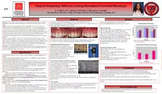
Fig. 1. Fig. 5: Papilla Height, by tooth type and phenotype. * Statistically significant. Fig 2. Papilla Height measurement. Fig. 3. Fig. 6: Average LID by phenotype. Fig. 4. Gingival Morphology Differences Among Determined Periodontal Phenotypes

Abstract
E N D
Presentation Transcript
Fig. 1 Fig. 5: Papilla Height, by tooth type and phenotype. * Statistically significant. Fig 2. Papilla Height measurement Fig. 3 Fig. 6: Average LID by phenotype Fig. 4 Gingival Morphology Differences Among Determined Periodontal Phenotypes L. N. MELL, H.-L. CHANG, P. KUMAR, T. SHI, and D.N. TATAKIS The Ohio State University, College of Dentistry, Division of Periodontology, Columbus, OH #33 Methods Abstract Results • 96 subjects, young adults averaging 24 yrs of age; female:male ratio was 73:23. • The study population was 82% non Hispanic Caucasians, 9% Asians, 6% African Americans, and two individuals listing themselves as “Other”. • Three phenotypes (A, B, C) were determined by cluster analysis (Fig. 1). • A: longer teeth, widest gingiva, thinnest gingiva • B: longest, most narrow crown form, narrowest gingiva, intermediate gingival thickness • C: shortest, most wide crown form, intermediate gingival width, thickest gingiva • Papilla Height (PH) Measurements • PH measurements were taken on casts. • PH measured from gingival line connecting zeniths of adjacent teeth. Measurement taken from the tip of the papilla to the line. • Measurements were parallel to long axis of corresponding medial tooth (Fig. 2). • Measurements were taken with a MH Ultra test electronic digital caliper to the nearest hundredth of a millimeter (Fig. 3). • Lateral Incisor Distance (LID) Measurements • LID = distance between gingival zenith of lateral incisor and determined gingival line. • LID measurements were completed on standardized gray scale photographs of casts (Fig. 4). • Gray scale photos were converted to black and white using Adobe Photoshop, and saved as TIFF files. • Scion imaging software was used to determine the gingival line between the zeniths of teeth #6 & #8 and to measure the LID. • Positive values were indicative of the lateral incisor zenith being coronal to the line. Negative values were indicative of the lateral incisor zenith being apical to the line. • Data Analysis • Descriptive statistics were calculated for mean (± SD) values and for frequency distributions. • One-way Analysis of Variance (ANOVA) was used to determine group mean differences, with post-hoc testing (Tukey-Kramer HSD) where appropriate. • Chi-square or Fisher Exact test were used as appropriate to test for frequency distribution differences. Objectives: The aim of this clinical investigation was to identify possible differences in gingival morphology between three periodontal phenotypes previously determined in a group of young adults. Methods: Systemically and periodontally healthy young adults (n=96; 18-36 years old) were recruited and three periodontal phenotypes (A, n=29; B, n=54; C, n=13) were determined using cluster analysis of 12 variables based on gingival width, thickness, and crown width/length ratio for teeth #7 and #8, as previously reported (Chang et al, IADR 2008, #2158). Subsequently, maxillary anterior dentition casts were used to measure height of interdental papilla (PH) for teeth #7 and #8. Lateral incisor distance (LID), the distance between the zenith of the lateral incisor and the line connecting the central incisor and canine zeniths (gingival line) was measured on standardized cast photographs. Differences in PH and LID between phenotypes were statistically analyzed. Results: The mean (± SD) PH values were 4.3±0.6 mm and 4.2±0.6 mm for teeth #7, and #8, respectively. Mean #8 PH was 3.9±0.6 mm, 4.4±0.5 mm, and 3.9±0.5 mm for phenotypes A, B, and C, respectively. #8 PH differed significantly between phenotypes (p<0.001), with phenotype B having the greatest #8 PH. In contrast, #7 PH did not differ significantly between phenotypes (p=0.161). The mean LID was 0.48±0.45 mm, with 5.5% of lateral incisor zeniths being apical to the gingival line. LID did not differ between phenotypes (p=0.233). Conclusions: The central incisor PH differs significantly between periodontal phenotypes, while the lateral incisor PH and LID do not. These results suggest that gingival morphology differences among periodontal phenotypes are site-specific. • Measurement reproducibility: the measurement error for both PH and LID measurements was less than 5%. Papilla Height (PH) • Mean (± SD) PH for the entire population was 4.2 ± 0.6 mm and 4.2 ± 0.6 mm for teeth #7 and #8, respectively. • Mean (±SD) #8 PH was 3.9 ± 0.6 mm, 4.4 ± 0.5 mm, and 3.9 ± 0.5 mm for phenotypes A, B, and C, respectively. #8 PH differed significantly between phenotypes (p < 0.0001), with phenotype B having the greatest #8 PH (Fig. 5). • Mean (±SD) #7 PH was 4.1 ± 0.6 mm, 4.3 ± 0.5 mm, and 4.2 ± 0.6 mm for phenotypes A, B, and C, respectively. #7 PH did not differ significantly between phenotypes (p=0.20) (Fig. 5). Lateral Incisor Distance (LID) • Mean (± SD) LID for the entire population was 0.48 ± 0.45 mm. • Mean LID was not significantly different between phenotypes (p=0.233) (Fig. 6). • 12% of zeniths were either flat (6.7%) with or apical (5.5%) to the gingival line. • The distribution of positive/negative zeniths did not differ between phenotypes (p=0.291). Introduction • Morphological characteristics of gingiva factor in diagnosis, treatment planning, and therapy. • Specifically, gingival morphology affects esthetics, susceptibility to trauma-induced recession, and periodontal surgical outcomes. • Different gingival phenotypes have been identified and characterized in the last decade. • Most phenotype studies limit characterization to gingival width and gingival thickness. • In a recent study from this institution, three periodontal phenotypes were identified in young adults (Chang et al 2008). The three phenotypes, A, B, and C, represented 30, 56, and 14% of the examined population, respectively. • For esthetic treatment planning, knowledge of other aspects of gingival morphology, such as interdental papilla height and relative position of gingival zenith, are also important. • There are no studies in the literature that have addressed such aspects of gingival morphology in relation to identified periodontal phenotypes. • The aim of this clinical investigation was to identify possible differences in specific gingival morphology between three periodontal phenotypes previously determined in a group of young adults. • The hypothesis was that interdental papilla height and lateral incisor gingival zenith position would be different among different phenotypes. • Specific aims: • Determine interdental papilla height (PH) for maxillary central (#8) and lateral (#7) incisors and statistically analyze PH differences between phenotypes. • Determine lateral incisor distance (LID), i.e., distance between the lateral incisor zenith and the line connecting the central incisor and canine zeniths (gingival line), and statistically analyze LID differences between phenotypes. Conclusions • Central incisor papilla height differed significantly between the three periodontal phenotypes. • Lateral incisor papilla height did not differ significantly between the three periodontal phenotypes. • Almost 90% of lateral incisor distance measurements were coronal to the gingival line connecting the zeniths between central and canine. • Lateral incisor distance measurements did not differ significantly between the three phenotypes. • These results suggest that particular gingival morphology differences between periodontal phenotypes are site-specific. Acknowledgements Supported by the Ohio State Division of Periodontology, the College of Dentistry, and the Ohio State University Barlow Dental Scholarship Fund


