Cytomegalovirus
470 likes | 663 Vues
Cytomegalovirus (CMV), a member of the beta-herpesvirus subfamily, is a double-stranded DNA virus that is ubiquitous in human populations. It can be transmitted vertically or horizontally, often without significant symptoms. In immunocompromised individuals, CMV can cause severe diseases such as pneumonia and retinitis. Congenital infections may lead to serious conditions like cytomegalic inclusion disease, affecting nearly 1% of live births. Diagnosis includes laboratory methods like PCR and antigen testing, while management varies based on the patient's immune status.

Cytomegalovirus
E N D
Presentation Transcript
Properties • Belong to the beta-herpesvirus subfamily of herpesviruses • ds DNA enveloped virus • Nucleocapsid 105 nm in diameter, 162 capsomers • Genome of CMV is similar to other herpesviruses, consisting of long and short segments which may be orientated in either direction, giving a total of 4 isomers. • A large no. of proteins are encoded for
Epidemiology • CMV is one of the most successful human pathogens, it can be transmitted vertically or horizontally usually with little effect on the host • Transmission may occur in utero, peri-natally or post-natally • Once infected, the person carries the virus for life which may be activated from time to time, during which infectious virions appear in the urine and the saliva.
Reactivation can lead to vertical transmission. It is also possible for people who have experienced primary infection to be re-infected with another or the same strain of CMV, this re-infection does not differ clinically from re-activation. • In developed countries with a high standard of hygiene, 40% of adolescents are infected and ultimately 70% of the population is infected. In developing countries, over 90% of people are ultimately infected
Pathogenesis • Once infected, the virus remains in the person for life and my be reactivated from time to time, especially in immunocompromised individuals. • The virus may be transmitted in utero, perinatally, or postnatally.
Perinatal infection: mainly through infected genital secretions, or breast milk. Overall, 2 - 10% of infants are infected by the age of 6 months worldwide. • Perinatal infection is thought to be 10 times more common than congenital infection. • Postnatal infection mainly occurs through saliva. Sexual transmission may occur as well as through blood and blood products and transplanted organ.
Clinical Manifestations • Congenital infection - may result in cytomegalic inclusion disease • Perinatal infection - usually asymptomatic • Postnatal infection - usually asymptomatic. However, in a minority of cases, the syndrome of infectious mononucleosis may develop which consists of fever, lymphadenopathy, and splenomegaly. • Heterophile Ab test is negative although atypical lymphocytes may be found in the blood.
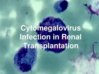
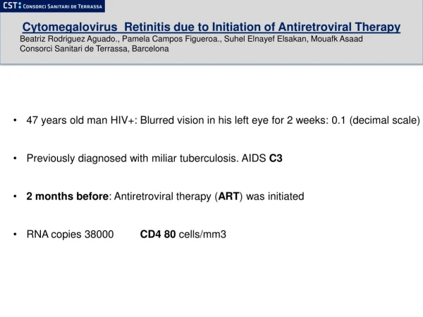
Immunocompromised patients such as transplant recipients and AIDS patients are prone to severe CMV disease: pneumonitis, retinitis, colitis, and encephalopathy • Reactivation or re-infection with CMV is usually asymptomatic except in immunocompromised patients.
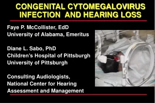
Congenital Infection • Isolation of CMV from saliva or urine within 3 weeks of birth. • Commonest congenital viral infection, affects 0.3 - 1% of all live births. • Second most common cause of mental handicap Responsible for more cases of congenital damage than rubella. • Transmission to fetus may occur following primary or recurrent CMV infection. 40% chance of transmission to fetus following a primary infection.
Cytomegalic Inclusion Disease • CNS abnormalities - microcephaly, mental retardation, spasticity, epilepsy, periventricular calcification • Eye - choroidoretinitis and optic atrophy • Ear - sensorineural deafness • Liver - hepatosplenomegaly and jaundice which is due to hepatitis.
Lung - pneumonitis • Heart - myocarditis • Thrombocytopenic purpura, Haemolytic anaemia • Late sequelae in individuals asymptomatic at birth - hearing defects and reduced intelligence.
Laboratory Diagnosis (1) • Direct detection • Biopsy specimens: examined histologically for CMV inclusion antibodies or for presence of CMV antigens. • pp65 CMV antigenaemia test is now routinely used for rapid diagnosis of CMV infection in immunocompromised patients. • PCR for CMV-DNA is used in some centers but there may be problems with interpretation.
CMV pp65 antigenaemia test (Virology Laboratory, New-Yale Haven Hospital)
Laboratory Diagnosis (2) • Virus Isolation: conventional cell culture is regarded gold standard but requires up to 4 weeks for result. • More useful are rapid culture methods such as the DEAFF test which can provide a result in 24-48 hours. • Serology: presence of CMV IgG antibody indicates past infection. Detection of IgM is indicative of primary infection, may be found in immunocompromised patients with reactivation.
Cytopathic Effect of CMV (Courtesy of Linda Stannard, University of Cape Town, S.A.)
DEAFF test for CMV (Virology Laboratory, New-Yale Haven Hospital)
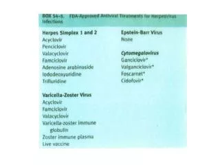
Treatment • Congenital infections - it is not usually possible to detect congenital infection unless the mother has symptoms of primary infection. If so, then the mother should be told of the chances of her baby having cytomegalic inclusion disease and perhaps offered the choice of an abortion. • Perinatal and postnatal infection - it is usually not necessary to treat such patients. • Immunocompromised patients - it is necessary to make a diagnosis of CMV infection early and give prompt antiviral therapy. Anti-CMV agents in current use are ganciclovir, forscarnet, and cidofovir.
Prevention • No licensed vaccine is available. There is a candidate live attenuated vaccine known as the Towne strain but there are concerns about administering a live vaccine which could become latent and reactivates. • Prevention of CMV disease in transplant recipients is a very complicated subject and varies from center to center. It may include the following measures. • Screening and matching the CMV status of the donor and recipient • Use of CMV negative blood for transfusions • Administration of CMV immunoglobulin to seronegative recipients prior to transplant • Give antiviral agents such as acyclovir and ganciclovir prophylactically.
Epstein-Barr Virus (EBV) • Belong to gamma-herpesvirus subfamily of herpesviruses • Nucleocapsid 100 nm in diameter, with 162 capsomers • Membrane is derived by budding of immature particles through cell membrane and is required for infectivity. • Genome is a linear double stranded DNA molecule with 172 kbp
The viral genome does not normally integrate into the cellular DNA but forms circular episomes which reside in the nucleus. • The genome is large enough to code for 100 - 200 proteins but only a few have been identified.
Epidemiology • Two epidemiological patterns are seen with EBV. • In developed countries, 2 peaks of infection are seen : the first in very young preschool children aged 1 - 6 • Second in adolescents and young adults aged 14 - 20 Eventually 80-90% of adults are infected. • In developing countries, infection occurs at a much earlier age so that by the age of two, 90% of children are sero-positive. • The virus is transmitted by contact with saliva, in particularly through kissing.
Pathogenesis • Once infected, a lifelong carrier state develops whereby a low grade infection is kept in check by the immune defenses. • Low grade virus replication and shedding can be demonstrated in the epithelial cells of the pharynx of all seropositive individuals. • EBV is able to immortalize B-lymphocytes in vitro and in vivo .
Furthermore a few EBV-immortalized B-cells can be demonstrated in the circulation which are continually cleared by immune surveillance mechanisms. • EBV is associated with several very different diseases where it may act directly or one of several co-factors
Disease Association 1. Infectious Mononucleosis 2. Burkitt's lymphoma 3. Nasopharyngeal carcinoma 4. Lymphoproliferative disease and lymphoma in the immunosuppressed. 5. X-linked lymphoproliferative syndrome 6. Chronic infectious mononucleosis 7. Oral leukoplakia in AIDS patients 8. Chronic interstitial pneumonitis in AIDS patients.
Infectious Mononuclosis • Primary EBV infection is usually subclinical in childhood. However in adolescents and adults, there is a 50% chance that the syndrome of infectious mononucleosis (IM) will develop. • IM is usually a self-limited disease which consists of fever, lymphadenopathy and splenomegaly. In some patients jaundice may be seen which is due to hepatitis. Atypical lymphocytes are present in the blood.
Complications occur rarely but may be serious e.g. splenic rupture, meningoencephalitis, pharyngeal obstruction. • In some patients, chronic IM may occur where eventually the patient dies of lympho proliferative disease or lymphoma. • Diagnosis of IM is usually made by the heterophile antibody test and/or detection of EBV IgM. • There is no specific treatment.
Burkitt’s Lymphoma (1) • Occurs endemically in parts of Africa (where it is the commonest childhood tumour) and Papua New Guinea. It usually occurs in children aged 3-14 years. It respond favorably to chemotherapy. • It is restricted to areas with holoendemic malaria. Therefore it appears that malaria infection is a cofactor. • Multiple copies of EBV genome and some EBV antigens can be found in BL cells and patients with BL have high titres of antibodies against various EBV antigens.
Burkitt’s Lymphoma (2) • BL cells show a reciprocal translocation between the long arm of chromosome 8 and chromosomes 14, 2 or 22. • This translocation result in the c-myc oncogene being transferred to the Immunoglobulin gene regions. This results in the deregulation of the c-myc gene. It is thought that this translocation is probably already present by the time of EBV infection and is not caused by EBV. • Sporadic cases of BL occur, especially in AIDS patients which may or may not be associated with EBV. • In theory BL can be controlled by the eradication of malaria (as has happened in Papua New Guinea) or vaccination against EBV.
Nasopharyngeal Carcinoma • Is a malignant tumour of squamous epithelium of nasopharynx. • Prevalent in S. China women. • Multiple copies of EBV genome and EBV EBNA-1 antigen can be found in cells of undifferentiated NPC. Patients with NPC have high titres of antibodies against various EBV antigens. • Environmental and genetic cofactors in NPC. • NPC usually presents late and thus the prognosis is poor.
Immunocompromised Patients • After primary infection, EBV maintains a steady low grade latent infection in the body. Should the person become immunocompromised, the virus will reactivate. In a few cases, lymphoproliferative lesions and lymphoma may develop. These lesions tend to be extranodal and in unusual sites such as the GI tract or the CNS. • Transplant recipients e.g. renal - EBV is associated with the development of lymphoproliferative disease and lymphoma. • AIDS patients - EBV is associated with oral leukoplakia and with various Non-Hodgekin’s lymphoma. • Ducan X-linked lymphoproliferative syndrome - this condition occurs exclusively in males who had inherited a defective gene in the X-chromosome . This condition accounts for half of the fatal cases of IM.
Diagnosis • Acute EBV infection: heterophil antibody test and/or detection of anti-EBV VCA IgM. • Cases of Burkitt’s lymphoma should be diagnosed by histology. The tumour can be stained with antibodies to lambda light chains which should reveal a monoclonal tumour of B-cell origin. In over 90% of cases, the cells express IgM at the cell surface. • Cases of NPC should be diagnosed by histology. • The determination of the titre of anti-EBV VCA IgA in screening for early lesions of NPC and also for monitoring treatment. • A patient with with non-specific ENT symptoms who have elevated titres of EBV IgA should be given a thorough examination.
Vaccination • Vaccine against EBV which prevents primary EBV infection should be able to control both BL and NPC. • Such a vaccine must be given early in life. Such a vaccine would also be useful in seronegative organ transplant recipients and those developing severe IM, such as the male offspring of X-linked proliferative syndrome carriers. • The vaccine should not preferably be a subunit vaccine since there is a danger that a live vaccine may still have tumorigenic properties. • The antigen chosen for vaccine development is the MA antigen gp 340/220 as antibodies against this antigen are virus neutralizing. • This vaccine is being tried in Africa.
Properties of HHV-6 and 7 • Belong to the beta-herpesvirus subfamily of herpesviruses • Double stranded DNA genome of 170 kbp • The main target cell is the T-lymphocyte, although B-lymphocytes may also be infected. • HHV-6 and HHV-7 share limited nucleotide homology and antigenic cross-reactivity. • It is thought that HHV-6 and HHV-7 are related to each other in a similar manner to HSV-1 and HSV-2.
Epidemiology and Pathogenesis • HHV-6 and HHV-7 are ubiquitous and are found worldwide. • They are transmitted mainly through contact with saliva and through breast feeding. • HHV-6 and HHV-7 infection are acquired rapidly after the age of 4 months when the effect of maternal antibody wears off. • By the time of adulthood, 90-99% of the population had been infected by both viruses. • Like other herpesviruses, HHV-6 and HHV-7 remains latent in the body after primary infection and reactivates from time to time.
Clinical Manifestations (1) • Primary HHV-6 infection is associated with Roseala Infantum, which is a classical disease of childhood. • Most cases: between the ages of 4 months and two years. • A spiking fever develops over a period of 2 days followed by a mild rash. The fever is high enough to cause febrile convulsions. • There are reports that the disease may be complicated by encephalitis.
Clinical Manifestations (2) • If primary infection is delayed until adulthood, there is a small chance that an infectious mononucleosis-like disease may develop in a similar manner to EBV and CMV. • There is no firm evidence linking HHV-6 to lymphomas or lymphoproliferative diseases. • There is no firm disease association with HHV-7 at present. • Although both viruses may be reactivated in immunocompromised patients, it is yet uncertain whether they cause significant disease since CMV is almost invariably present.
Diagnosis and Management • Rosela Infantum has a very characteristic presentation and a diagnosis can usually be made on clinical grounds alone. • Therefore very few virology laboratories offer a diagnostic service for HHV-6 or HHV-7 infection. • The technique for virus isolation is complicated and thus not practicable as a routine diagnostic procedure. • Therefore serology is the mainstay of diagnosis where specific IgM and IgG are detected. • There is no specific antiviral treatment for HHV-6 infection.
Human Herpes Virus 8 • Belong to the gammaherpesviruses subfamily of herpesviruses • Originally isolated from cells of Kaposi’s sarcoma (KS) • Now appears to be firmly associated with Kaposi’s sarcoma as well as some lesser known malignancies such as Castleman’s disease and primary effusion lymphomas • HHV-8 DNA is found in almost 100% of cases of Kaposi’s sarcoma
Most patients with KS have antibodies against HHV-8 • The seroprevalence of HHV-8 is low among the general population but is high in groups of individuals susceptible to KS, such as homosexuals. • Unlike other herpesviruses, HHV-8 does not have a ubiquitous distribution.