Download
1 / 16
260 likes | 930 Vues
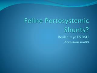
Feline Portosystemic Shunts?. Beulah, 2 yo FS DSH Accession 110188. Beulah. 2 yo FS DSH with behavioral change towards extreme aggression 3 seizure-like episodes, beginning at 6 months of age Physical exam performed under general anesthesia: unremarkable Lesion localization: forebrain

E N D
Feline Portosystemic Shunts? Beulah, 2 yo FS DSH Accession 110188
Beulah • 2 yo FS DSH with behavioral change towards extreme aggression • 3 seizure-like episodes, beginning at 6 months of age • Physical exam performed under general anesthesia: unremarkable • Lesion localization: forebrain • MRI findings reputedly consistent with a portosystemic shunt • CSF unremarkable • Ammonia levels: 52 and 58 umol/L on two separate occasions • Resting bile acids: 7.6 umol/L
BRAIN MRI CHARACTERISTICS IN DOGSAND CATS WITH CONGENITAL PSS SHUNTS • All animals with PSS had widened sulci, esp large breed dogs • 10/13 dogs and 1/3 cats had hyperintensity of the lentiform nuclei on T1W images, extend outward to involve the nucleus accumbens that did not contrast enhance, was not present on T2W images • Proposed causes: lipid deposition, calcification, • melanin deposition, methemoglobin production, or manganese (Mn) accumulation • After surgical ligation, one dog and one cat were re-imaged, with a decrease in the lentiform nuclear hyperintensity
Feline, pre- and post-ligation Canine w/ PSS pre-ligation T1W, T2W Canine w/ PSS Pre-ligation T1 W, transverse and dorsal planes
Manganese • Found in human patients with cirrhosis and similar findings? • Elevated in blood with TPN administration • Mn2+ is paragmagnetic: shortens T1
Lentiform nuclei • Part of the basal nuclei; grey matter of the telencephalon • Includes the globuspallidus and putamen • Telencephalon Function • Connections with cerebrum and thalamus • Cortex inhibitory to BN • Cortical lesions – hyperactivity • BN lesions – hypoactivity • Motor activity • Planning, execution • Humans • More important
Beulah’s Ultrasound? • Normal liver size, with normal portal conspicuity • Renal size: 42 mm and 45 mm • No urinary calculi • No evidence of a portosystemic shunt noted • PV=0.375 cm • PV/CVC=0.70
Feline portosystemic shunts • Typically a single, large anomalous communication between the abdominal veins that arise from the vitelline veins and the cardinal veins • The right vitelline vein forms the first tract of the caudal vena cava • and portal vein • The cranial portion of left umbilical vein becomes the Aranzioductusvenosus; • The right umbilical vein disappears (right vitelline to right cardinal anastomoses are source of EHPSS) • From the cardinal vein system the azygos vein and caudal pre-renal vena cava originate. • Between the second and the sixth day of the life ductusvenosuscloses. • Extrahepatic shunts make up 90% of the feline congenital PSS cases • Normal feline PV arises from mesenteric vein, gastrosplenic vein, gastroduodenal and cranial pancreaticoduodenal • Supplies 75-80% of blood supply to liver with portal pressures of 10-13 mm Hg • Congenital PSS may shunt up to 80% of the normal portal blood flow away from the liver
Feline PSS • Breed: largest group=DSH, but Persian, Siamese, Himalayan, and Burmese are frequent • Hard to palpate liver, prominent kidneys • Copper-colored irises • Congenital cardiac murmurs, a peritoneopericardial hernia, and a high rate of cryptorchism (24%) reported • Hepatic encephalopathy: altered consciousness, behavioral changes, visual deficits (amaurosis), ataxia, head pressing, circling, etc • GI signs including ptyalism • PU/PD, lower urinary tract signs, ammonium biuratecrystalluria
Feline PSS • Blood work similar to dog, but less consistent • Elevated bile acids, and elevated ammonia levels (because hepatocyte conversion of ammonia to urea is reduced), elevation in ammonia levels with ATT, ammonia tolerance test • Transcolonic portal scintigraphy shunt fractions: dogs average 84%, cats average 52% (normal in cat is 5.9%)
The US diagnosis of PSS • In both dogs and cats: • 92% sensitive, 98% specific • PPV=98%, NPV=89%, • Overall accuracy of 95% • PV/Ao <0.65: extrahepatic PSS or idiopathic noncirrhotic portal hypertension • PV/Ao > 0.8 and PV/CVC >0.75, respectively, did not have an extrahepatic PSS • Turbulence in the CVC in DOGS • PPV=91% and NPV=84%,
US diagnosis of PSS-feline details • Only 22% of cats with PSS had a small liver • 17.1 cm/s (9.7–18.1) were portal velocities for normal cats previous reports 10-12 cm/s • Portal vein diameter may be as accurate as ratios in cats, as the diameter does not vary with size • No PSS: PV=0.44 cm (0.34-0.5 cm) • PSS: PV=0.27 cm (0.20-0.35 cm) • Renomegaly and decreased portal conspicuity are less commonly seen in felines
PORTOGRAPHY • VD and Llat, or Llat alone with mesenteric portography • EHPSS will have no opacification of the liver • IHPSS may have partial opacification • Most common PSS in cats arises from the left gastric vein or the portal vein and drains into the caudal vena cava (portocaval shunt) between the phrenicoabdominal vein and the liver • Second most common: portoazygous • Location more variable in cats than dogs
Feline PSS • Medical management (similar to dogs): protein restriction, aggressive tx of gastric ulceration, lactulose, antibiotics to reduce colonic flora • Surgical management: • Normal 10-13 cm H2O • Post-ligation recommendations: • Never over 20 cm H2O • Never rise >10 cm H2O • 34% of cats may undergo complete ligation based on these guidelines, with second surgery to follow for complete ligation • Reported mortality ranges from 10-71% • Complications: hemorrhage, fatal portal hypertension, ascites, seizures • Prognosis good with complete ligation, poorer than dogs with partial ligation
References • D’Anjou MA, et al. Ultrasonographic diagnosis of portosystemic shunting in dogs and cats. Veterinary Radiology & Ultrasound, Vol. 45, No. 5, 2004, pp 424–437. • Santilli RA and Garboni G. Diagnostic imaging of congenital porto-systemic shunts in dogsand cats: a review. The Veterinary Journal 166, 2003, pp 7–18. • Tillson DM, Winkler JT. Diagnosis and treatment of portosystemic shunts in the cat. Clin Small Anim 32, 2002, pp 881–899. • Torisu, et al. Brain magnetic resonance imaging characteristics in dogs and cats with congenital portosystemic shunts. Veterinary Radiology & Ultrasound, Vol. 46, No. 6, 2005, pp 447–451. • Tidwell. Neuroanatomy slides