Download
1 / 46
490 likes | 1.04k Vues
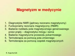
Magnetyzm w medycynie. Diagnostyka NMR (jądrowy rezonans magnetyczny) Funkcjonalny rezonans magnetyczny (fNMR) Badanie rozkładu pola magnetycznego generowanego przez prądy – diagnostyka mózgu i serca Badanie magnetyczne przewodu pokarmowego Termoterapia za pomocą pola zmiennego

E N D
Magnetyzm w medycynie • Diagnostyka NMR (jądrowy rezonans magnetyczny) • Funkcjonalny rezonans magnetyczny (fNMR) • Badanie rozkładu pola magnetycznego generowanego przez prądy – diagnostyka mózgu i serca • Badanie magnetyczne przewodu pokarmowego • Termoterapia za pomocą pola zmiennego • Termoterapia za pomocą cząstek magnetycznych B. Augustyniak
Natężenia pola magnetycznego w środowisku Natężenia pola magnetycznego dla różnych źródeł oraz zakresy czułości mierników pola Magnetism in Medicine – Handbook; WILEY-VCH Verlag GmbH, Weinheim, 2007 B. Augustyniak
Biomagnetyczne efekty Bioelectromagnetic interactions and phenomena as a function of their magnetic flux and frequency range. Recent Advances in Biomagnetics and Bioimaging for Brain Research and Sensing Technologies, IEEE SENSORS 2009 Conference B. Augustyniak
Właściwości magnetyczne tkanek Spectrum of magnetic susceptibilities. The figure shows thatthe majority of human tissues is diamagnetic or weakly paramagnetic.. http://www.biomedical-engineering-online.com/content/3/1/11 B. Augustyniak
Właściwości elektryczne tkanek Frequency dependence of relative permittivity ε (x) and conductivityσ (o); the major dispersion regions α, β and γ areindicated. The frequency range of interest for MRI devices isreported. http://www.biomedical-engineering-online.com/content/3/1/11 B. Augustyniak
Przewodnictwo elektryczne mózgu Images of the Mean Conductivity (MC) averaged on the threedirections for the motion-probing gradients (anterior-posterior, right-left, andsuperior-inferior), and the Anisotropy Index (AI), an index of the degree oftissue anisotropy in conductivity. Detection of weak magnetic fields arising from neuronalelectrical activities using MRI is a potentially effectivemethod for functional imaging of the human brain (Figure above).We have measured the transient changes in MR signalintensity arising from neuronal electrical activities. Recent Advances in Biomagnetics and Bioimaging for Brain Research and Sensing Technologies, IEEE SENSORS 2009 Conference B. Augustyniak
Historia NMR nobliści 1991chemia wkład do rozwoju metodologii NMR o dużej rozdzielczości Richard R. Ernst Felix. Bloch Edward Purcell 1952fizyka rozwój nowych precyzyjnych metod pomiarów magnetyzmu jądrowego 2003medycyna odkrycia dotyczące obrazowania za pomocą rezonansu magnetycznego Paul C. Lauterbur Sir Peter Mansfield B. Augustyniak
Momenty magnetyczne jąder w stałym polu B B = 0 precesja pole B B >0 Częstość precesji Larmoraw = g B Dla wodoru: fL [ MHz ] = 42,6 Bo [ T ]
Przekrój tomografu RM Simens 3T Alegra B. Augustyniak
Głowa i szyja Kręgosłup szyjny i piersiowy Piersi Miednica Kończyny Serce, płuca, brzuch Przykłady cewek nadawczo-odbiorczych amu.edu.pl/~ewas/pracownia/img/Tomografia%20NMR.ppt B. Augustyniak
Układ blokowy aparatu NMR NS – magnes główny, A – cewka nadawczo-odbiorcza, U – komutator, G – nadajnik, R – odbiornik, K – układ sterowania i analizy sygnałów B. Augustyniak
z Pomiar czasów relaksacji dla tkanek y Bo wzbudzanie: impuls RF90o x y . . z x Wirujące pole o składowej MT Cewka wzbudzająca impuls RF y x z . . FID Detekcja: Indukowane napięcie w tej cewce na skutek zmiany składowej MT
Pole magnetyczne zmienne generowane w tomografie RM Zmienne pola elektromagnetyczne emitowane przez tomograf RM (3ms/dz) Praca naukowo-badawcza z zakresu prewencji wypadkowej, CIOP, 2008 B. Augustyniak
Obrazy NMR -3 bark głowa kolano wątroba
Zobaczyć w kolorach wykorzystać T1, T2 i gęstość r protonów
Zagrożenia w badaniach NMR • Pole stałe magnetyczne 2. Pole zmienne magnetyczne B. Augustyniak
Przeciwwskazania i ryzyko metody • Silne stałe pole magnetyczne – nieszkodliwe do wartości 2 T (Bezwzględne przeciwwskazania – stymulatory pracy serca i metalowe implanty) • Gradienty pola magnetycznego i ich przełączanie – prądy indukowane mogą powodować ogrzewanie organizmu, powyżej 6 T/s • Efekty akustyczne przełączania gradientów – rzędu 65 – 95 dB, zalecane stopery do uszu. W nowoczesnych tomografach – efekty dźwiękowe nie są dokuczliwe B. Augustyniak amu.edu.pl/~ewas/pracownia/img/Tomografia%20NMR.ppt
Pole magnetyczne w pobliżu tomografu RM B. Augustyniak Praca naukowo-badawcza z zakresu prewencji wypadkowej, CIOP, 2008
Strefy zagrożeń wokół źródła pola B. Augustyniak Praca naukowo-badawcza z zakresu prewencji wypadkowej, CIOP, 2008
Strefy ochronne w pobliżu tomografu RM B. Augustyniak Praca naukowo-badawcza z zakresu prewencji wypadkowej, CIOP, 2008
Granice stref ochronnych dla MR Natężenia pola magnetycznego na granicy stref ochronnych w odniesieniu do częstotliwości wykorzystywanych w tomografach RM B. Augustyniak Praca naukowo-badawcza z zakresu prewencji wypadkowej, CIOP, 2008
fMRI Funkcjonalny rezonans magnetyczny fMRI MRI (NMR) Functional Magnetic Resonance Imaging http://psychology.uwo.ca/fMRI4Newbies/Tutorials/01%20Intro/Introduction.ppt Bolesław AUGUSTYNIAK
fMRI schemat aparatury Bolesław AUGUSTYNIAKfMRI [2]
Hemoglobin Source: http://wsrv.clas.virginia.edu/~rjh9u/hemoglob.html, Jorge Jovicich Hemoglogin (Hgb): - four globin chains - each globin chain contains a heme group - at center of each heme group is an iron atom (Fe) - each heme group can attach an oxygen atom (O2) - oxy-Hgb (four O2) is diamagnetic no B effects - deoxy-Hgb is paramagnetic if [deoxy-Hgb] local B Bolesław AUGUSTYNIAKfMRI
BOLD kontrast Blood Oxygenation Level Dependent (BOLD) contrast, because the amount of deoxyhemoglobin depends onthe oxygen saturation of the blood. At rest, approximately 40% of the cortical blood volume is deoxygenated. Thesmall rise in total oxygen extraction following neuronal activation is overcompensatedby oxygen delivery due to the rise in blood flow. Therefore, thelevel of deoxyhemoglobin is reduced to approximately 20–25%, and this reductionleads to a signal rise during neuronal activation.
BOLD signal Blood OxygenLevelDependent signal • neural activity blood flow oxyhemoglobin T2* MR signal Mxy Signal Mo sin T2* task T2* control Stask S Scontrol time TEoptimum Source: fMRIB Brief Introduction to fMRI Source: Jorge Jovicich http://www.mntp.pitt.edu/Workshop/MNTP_res_2008/MNTP_fMRI_history_Kim.ppt Bolesław AUGUSTYNIAK
2. Category-Specific Visual Areas objects faces Malach, 2002, TICS • Parahippocampal Place Area (PPA) • place-selective • places > (objects and faces) • places > scrambled images places • Lateral Occipital (LO) • object-selective • objects > (faces & scenes) • objects > scrambled images • Fusiform Face Area (FFA) or pFs • face-selective • faces > (objects & scenes) • faces > scrambled images • ~ posterior fusiform sulcus (pFs) http://psychology.uwo.ca/fMRI4Newbies/Tutorials/01%20Intro/Introduction.ppt Bolesław AUGUSTYNIAK
Badanie aktywności mózgu metodą fNMR (a) Flat map representation of the activation in thehuman motion-sensitive area V5/V5a during a visual motionperception (moving concentric rings, right above). (b) A rendered head volume during a visual stimulus Magnetism in Medicine – Handbook; WILEY-VCH Verlag GmbH, Weinheim, 2007 B. Augustyniak
Badania rozkładów pola magnetycznego generowanego przez prądy płynące lokalnie w ciele www.mp.uni-tuebingen.de/mp/uploads/media/Christoph_Braun.pdf B. Augustyniak
Badanie pola magnetycznego mózgu Cutaway drawing of the „Magnes” dewar showing thelocation of the reference channels relative to the sensor coils Magnes 3600 WH in position for a seated study. Magnetism in Medicine – Handbook; WILEY-VCH Verlag GmbH, Weinheim, 2007 B. Augustyniak
Sygnały z czujników pola magnetycznego Waveform, field distribution and sourcelocalization data for 13-month-old infant with infantile spasms studied with conscioussedation. The patient was positioned with herhead centered in the sensor array of a Magnes 2500 WH 148-channel system. Magnetism in Medicine – Handbook; WILEY-VCH Verlag GmbH, Weinheim, 2007 B. Augustyniak
Badanie akcji mózgu systemem VSM MedTech (a) Cortical 275-channel CTF MEGTM System (VSM MedTech).(b) MEG of a somatosensory-evoked field recorded by CTF MEGTM System (DC to 300 Hz bandwidth, 628 averagesand third-order gradiometer noise cancellation). Magnetism in Medicine – Handbook; WILEY-VCH Verlag GmbH, Weinheim, 2007 B. Augustyniak
Lokalizacja ‘muzyki’ Left:(MEG-based brainsurface current density (BSCD) reconstructions of motor activity inmusicians listening to piano music (Haueisen and Knosche, 2001). Magnetism in Medicine – Handbook; WILEY-VCH Verlag GmbH, Weinheim, 2007 B. Augustyniak
Badania magnetyczne pracy serca • Locus of the total current dipole vector, P, duringthecardiaccycle and magnetocardiogram in a healthy subject. • Signal-averaged traces from a 61-channel prethoracic acquisition. • Magnetic field map at Q onset schematically superimposed on anMR image. Magnetism in Medicine – Handbook; WILEY-VCH Verlag GmbH, Weinheim, 2007 B. Augustyniak
Diagnostyka magnetyczna przewodu pokarmowegoza pomocą śledzenia ruchu „magnetycznych” elementów
Badania magnetyczne pracy przewodu pokarmowego PTB 83-SQUID system. (a) Path of the magnetically marked dosage form throughthe GI tract of a volunteer in the fasted state. PTB 83-SQUID system. (a)system inside the shielded room SQUID, (b) S configuration (top view); Magnetism in Medicine – Handbook; WILEY-VCH Verlag GmbH, Weinheim, 2007 B. Augustyniak
Thermoterapia magnetyczna Wykorzystanie prądów wirowych Wykorzystanie cząstek magnetycznych
1. Magnetoterapia za pomocą zmiennego pola magnetycznego Generator prądu (a) i aplikatory (b) (cewki szpulowe) przykładowego urządzenia do magnetoterapii B. Augustyniak Praca naukowo-badawcza z zakresu prewencji wypadkowej, CIOP, 2008
Thermoterapia z wykorzystaniem ogrzewania cząsteczek magnetycznych wymuszonego zmiennym polem magnetycznym
2. Termoterapia za pomocą cząsteczek magnetycznych Frequency dependence of temperature rise for ferritepowders after an application of AC magnetic field for 2 min: Core loss for ferrite powder as function of frequency atroom temperature ð25CÞ under HAC ¼ 30 A=m: Nanoparticles in photodynamic therapy: An emerging paradigm; Advanced Drug Delivery Reviews Volume: 60, Issue: 15, December 14, 2008, pp. 1627-1637; Chatterjee, Dev Kumar; Fong, Li Shan; Zhang, Yong B. Augustyniak
Termoterapia za pomocą pola f =100 kHz i płynów magnetycznych Sketch of the first prototype MFH therapy system (MFH Hyperthermiesysteme GmbH,Berlin,Germany). The AC magnetic field axis is perpendicular to the axial direction of the patient couch (1). The therapy system is for universal application,i.e.,suitable forMFH within,in principle,any body region. It is a ferrite-core applicator (2) operating at a frequency of 100 kHz with an adjustablevertical aperture of 30-50 cm (3). The field strength is adjustable from 0 to 15 kA/m. The system is air cooled (4). Presentation of a new magnetic field therapy system for the treatment of human solid tumors with magnetic fluid hyperthermia, Journal of Magnetism and Magnetic Materials Volume: 225, Issue: 1-2, 2001, pp. 118-126 Jordan, A.; at all B. Augustyniak
Termoterapia za pomocą pola zmiennego 3D field strength distributions were calculated using the ‘finite-difference-time-domain-(FDTD)-method‘ . One example of the visualized results is shown in Fig. 3A (3Dvector diagram,coronary view,24 cm aperture, 15 kA/m) Presentation of a new magnetic field therapy system for the treatment of human solid tumors with magnetic fluid hyperthermia; Journal of Magnetism and Magnetic Materials Volume: 225, Issue: 1-2, 2001, pp. 118-126 Jordan, A et. al. B. Augustyniak
Zwalczanie raka Presentation of a new magnetic field therapy system for the treatment of human solid tumors with magnetic fluid hyperthermia; Journal of Magnetism and Magnetic Materials Volume: 225, Issue: 1-2, 2001, pp. 118-126 Jordan, A et. al. B. Augustyniak