Plant Biology Fall 2006
360 likes | 634 Vues
Plant Biology Fall 2006. BISC 367 - Plant Physiology Lab Spring 2009. 1.0. Plant Physiology Lab Spring 2009 Professor: Dr. Aine Plant, office B8228 e-mail: aplant@sfu.ca (preferred) Tel: 778-782-4461 Lab Instructor: Doug Wilson, office B9239

Plant Biology Fall 2006
E N D
Presentation Transcript
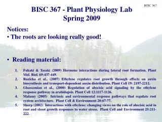
Plant Biology Fall 2006 BISC 367 - Plant Physiology Lab Spring 2009
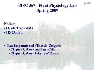
1.0 Plant Physiology Lab Spring 2009 Professor: Dr. Aine Plant, office B8228 e-mail: aplant@sfu.ca (preferred) Tel: 778-782-4461 Lab Instructor: Doug Wilson, office B9239 e-mail: dwilson@sfu.ca TA: Owen Wally e-mail: owenw@sfu.ca Lectures: Tuesday at 11:30 - 12:20 AQ 4120 Lab & tutorial: Thursday 1:30 - 5:20 in B8241 Thursday 11:30 to 12:20 in B8241 (not in AQ5049)
1.1 Mark distribution: 2 quizzes 10 % each 2 lab reports 17.5 % each Lab report based on project 25 % Lab worksheets 20% Quiz 1: Tuesday Feb. 10 Quiz 2: Tuesday Mar. 24 Project report due: First week of exams Textbook: Taiz and Zeiger “Plant Physiology” 4th edition On reserve in the library
1.1 Online material: http://www.sfu.ca/bisc/bisc367/ • Course outline • Lab handouts • Posted lecture presentations • Lab project data and info.
1.1 Plant Physiology Lab Spring 2009 • Notices: • General reading: • Chapter one focus on: • Tissues • Chloroplasts • Plasmodesmata • Chapter 15 covers cell walls. Cover the basics only!
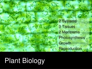
Overview - plant morphology • Shoot system • Stem • Supports and places leaves • Transports H2O and nutrients • Leaves • Photosynthesizers • Reproductive structures • Root system • Anchors plant • Absorbs water and minerals • Storage (CHO) & synthesis of some hormones
Overview - plant morphology • 3 major tissue systems make up the plant body • Ground tissue • cortex • mesophyll • pith • Vascular tissue • Dermal tissue • Tissue systems are continuous throughout the plant
3 Tissue Systems • Ground tissue includes: • Parenchyma tissue • Collenchyma tissue • Sclerenchyma tissue • Vascular tissue includes • Xylem tissue • Phloem tissue • Dermal tissue • Epidermis
Tissue Systems • Parenchyma tissue: • SIMPLE • Made up of a single cell type • Cells are ALIVE at maturity • Capable of dividing • TOTIPOTENT • Involved in wound regeneration and range of metabolic fxns
Tissue Systems • Chollenchyma tissue: • SIMPLE • Cells are ALIVE at maturity • Contain unevenly thickened walls • Support young growing stems and organs
Tissue Systems • Sclerenchyma tissue: • SIMPLE • Cells are dead at maturity • Typically lack protoplasts • Possess secondary walls with lignin • Strong polymer • Support stems and organs that have stopped growing fibres sclereid Economically important tissue! e.g. Hemp fibres
Tissue Systems • Xylem tissue: • COMPLEX • Made up from more than one cell type • Functions • Conduction of H2O • Structural support • Cells are elongated & dead at maturity • Lack protoplasts • Possess elaborately thickened secondary walls with lignin (very strong) • 2 main cell types • Vessel members • Tracheids
How does H2O pass from one tracheid to the next? • Passes through aligned pits of neighbouring tracheids • Pit membrane consists of 1o wall only Tracheid 1 Pits Tracheid 2 Tissue Systems • Tracheids (primitive): • Tracheids “stack” longitudinally in the stem overlapping at tapered ends
3 vessel members stacked end to end to form part of a vessel Tissue Systems • Vessel members (advanced): • Stack end to end to form a vessel (long) • Perforation plate at ea. end of a member permits easy water flow Slotted perforation plate forms end wall of a vessel member Water passes from vessel to vessel via pits
Tissue Systems • Xylem is a complex tissue: • Also present • Parenchyma tissue (nutrient storage) • Fibres/sclereids
Tissue Systems • Phloem tissue • Complex • Functions • Conduction of nutrients • Cells are alive at maturity but highly modified • Lack: • Nucleus • Definition between cytoplasm and vacuole • 2 main cell types • Sieve cells • Sieve tube members
Sieve tube member 1 Sieve plate Sieve tube member 2 Tissue Systems • Sieve tube members (advanced) • Elongated cells • Sieve tube members stack end to end to form a sieve tube • End walls form sieve plates and contain pores that connect the the cytoplasm of two sieve cells for solute transfer
Tissue Systems • Sieve tube members and sieve cells are connected to specialized cells • A sieve tube member is always associated with a companion cell • Connected via plasmodesmata • companion cell provides: • metabolic functions • Loads sugars for transport
Waxy cuticle Tissue Systems • Dermal tissue • Functions • Mechanical protection • Made up of epidermal (parenchymal) cells • Cells overlaid with a waxy cuticle to minimize H2O loss
Stomatal pore Tissue Systems Dermal tissue • Also present • Guard cells • Regulate size of the stomatal pore and • Movement CO2 into leaf • Movement H2O vapour out
Branched & glandular trichomes Tissue Systems Dermal tissue • Also present • Trichomes aka “hairs” • Increase reflectance of solar radiation • Absorb H2O and minerals (epiphytes) • Contain chemical defenses • Can impale larvae of some insects
Root epidermal cell with root hair Root anatomy • Root structure • Simple • Epidermis (outer layer of cells) • Protects root • Plays important role in water uptake • Facilitated by root hairs • Tubular extension from epidermal cell • Increases surface area for water uptake • Produced in zone of maturation • Short lived
Root anatomy • Cortex • Ground tissue that occupies most volume of root • Cells often adapted for storage • Starch • Numerous air spaces exist • Roots need to respire! • Innermost boundary of cortex is the endodermis
Root anatomy • Vasculature in a eudicot root • Protostele • Vascular tissue occupies the centre of root • Xylem arranged as a “star” • Phloem tissue is located between the arms of the xylem “star” • Pericycle tissue surrounds vascular tissue
Central pith Maize root Root anatomy • Vasculature in some monocot roots develops with a central pith
Stem anatomy • Primary structure of a eudicot stem • 1o vascular tissue are present as a cylinder of strands separated by ground tissue • Interfascicular rays or pith rays • 1o phloem is present at the outside of the bundle • 1o xylem is present on the inside of the bundle • Ground tissue in centre of stem is the pith • Ground tissue that lies outside the vascular bundle is the cortex • Outermost layer is the epidermis • Contains stomata and trichomes
Stem anatomy • Primary structure of a eudicot stem • Single layer of cells between 1o phloem & 1o xylem remain meristematic • Become vascular cambium • Cylindrical meristem that is responsible for 2o growth • Remainder of cambium arises from interfascicular parenchyma • Note, not all eudicots undergo 2o growth • No cambium arises
Anatomy of a woody stem • Woody stem during first year of growth
Leaves • Evolved to photosynthesize • Divided into • Blade or lamina • Petiole or stalk • Leaf anatomy is influenced by the amount of available water: • Plants can be grouped according to their water requirements: • mesophyte • Plant with plentiful water supply • hydrophyte • Grows partially or completely submerged • xerophyte • Adapated to dry environment
Leaf anatomy • General features of mesophytic leaves (eudicot) • Stomata more numerous on lower surface • sheltered • Photosynthetic tissue (mesophyll) is differentiated into: • Upper palisade parenchyma • Upright cells with many cps • Lower spongy mesophyll • Permeated by air spaces • Vasculature is netted venation • Xylem towards upper surface • Phloem towards lower surface • Small veins collect P/S products • Surrounded by a bundle sheath • Controls entry/exit of material • Large veins transport P/S products from leaf
Leaf anatomy • Anatomical modifications in hydrophytes • Problem = obtaining enough CO2 & O2 • Stomates not present or in upper epidermis (floating leaf) • Thin cuticle • Large amounts of air in spongy mesophyll • Gas exchange • buoyancy • Reduced vascular tissue • Partic. xylem • Reduced amount of support tissue
Leaf anatomy Modifications present in xerophytes • Problem = getting enough water • Many of these plants have reduced leaf size or no leaves • Large number of stomates • Optimize gas exchange when water is plentiful? • Remember stomates usually shut • Stomates generally sunk in depression in leaf surface • Assoc. with trichomes • Both increase depth of boundary layer & slow rate of water loss • Thick cuticle • Multiple epidermis • Modified to store water • More supporting tissue to compensate for reduced turgor Stomate
P/S CO2 + C3 acid CO2 + C3 acid C4 acid C4 acid Mesophyll cell Bundle sheath cell Leaf anatomy • General features of monocot leaves • Parallel venation system • Lack a defined palisade/spongy mesophyll layers • Leaves tend to be vertically oriented • Anatomy modified according to mode of P/S • C4 photosynthesis • Carbon fixed to form a C4 acid in mesophyll cell • C4 acid is transported to bundle sheath cell & decarboxylated • Released CO2 is refixed by C3 P/S
Leaf anatomy • Leaves of C4 plants display Kranz anatomy • Mesophyll and BSC form 2 concentric layers around a vascular bundle • Bundle sheaths are close together • Leaves of C3 plants have well separated bundle sheaths and do not have Kranz anatomy C4 leaf C3 leaf