Ocular trauma
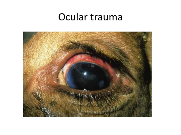
Ocular trauma. Clinical features:. although the eye is well protected within the bony orbit and by the rapid reflex closure of the lids to approaching foreign bodies, traumatic eye lesions are common, particularly those due to incoming objects.

Ocular trauma
E N D
Presentation Transcript
Clinical features: • although the eye is well protected within the bony orbit and by the rapid reflex closure of the lids to approaching foreign bodies, • traumatic eye lesions are common, particularly those due to incoming objects. • Irritation due to dust or ultraviolet light may produce keratitis and conjunctivitis. • Management: • most cases resolve completely within a few days. If persistent, a further check for a foreign body should be made before starting topical therapy.
Clinical features: • grass seeds or other plant material may become lodged in the conjunctiva • as the eyeball moves, repeatedly traumatize the area to produce erosion and ulceration. • Cattle reaching up to feed from overhead hay racks are particularly at risk. • Management: • following good restraint and possibly use of a topical local anesthetic, the foreign body can be removed manually or with fine, blunt-tipped forceps. • Topical therapy should be given for several days. • Small foreign bodies that become embedded in the cornea • and produce a keratitis will often resolve spontaneously over time.
Definition: • forward displacement or bulging of the eye. • Clinical features: • an infrequent condition caused by trauma to the head. Management: • under sedation and local anesthesia • the eyeball can be returned to its socket. • If held in placefor4–5 days by suturing the lids together, most cases resolve well.
Clinical features: • lacerations of the lower eyelids are fairly common. • They are often caused by an animal rubbing the head and catching an eyelid on projections from troughs, buildings, or fragments of wire. In the • Management: • sometimes the resultant rough eyelid edge leads to incomplete lid closure with persistent lowgradecorneal ulceration and lacrimation. More severe cases should therefore benefit from suturing.
Pathogenesis: • squamous cell carcinoma (SCC) is the most common ocular neoplasm of cattle, and is seen particularly in mature white-headed beef cattle, such as the Hereford, and other breeds with little pigmentation around the eye (e.g., Simmental). The disease is associ-atedwith sunlight (ultraviolet light). • Clinical features: • common sites for SCC include the lower lid, the third eyelid, and the corneoscleraljunction of the globe. Often both eyes are affected to a varying degree. Small benign precursor lesions will often regress. infection. If neglected, about 10% of cases will eventually metastasize to the regional lymph nodes (resulting in slaughterhouse condemnation
Differential diagnosis: • IBK , • lymphosarcoma . • Management: • early third eyelid lesions are easily • removed under sedation and local anesthesia using • forceps and scissors. No suturing is required. More advanced cases require cryo-therapy or total removal of the eyeball to avoid regional spread.
a papilloma is attached to the third eyelid by a “stalk” and has a very irregular keratinized surface. It is much less common than an SCC, and is easy to remove surgically. • Periocularpapillomata (“warts”) are frequently seen in groups of younger calves. • three mature papillomata originating from the skin of the eyelids at medial and lateral canthus. Several small papillomata can be seen distant from the eye.