Dynamics of Electropore Formation in Time-Dependent Electric Fields
This study investigates the effects of time-dependent electric fields on the electroporation of cell membranes, utilizing molecular dynamics simulations. By varying frequencies, amplitudes, and pulse shapes, we show that pore properties can be dynamically modulated, providing insights that refine existing electroporation models. Our results showcase how external electric fields can hold pores open and control their expansion, which may pave the way for advanced therapeutic and engineering applications in cellular manipulation. This work presents a unified framework for future studies in electroporation.

Dynamics of Electropore Formation in Time-Dependent Electric Fields
E N D
Presentation Transcript
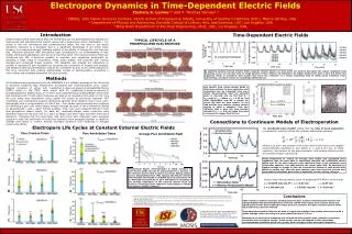
Electropore Dynamics in Time-Dependent Electric Fields Zachary A. Levine1,2 and P. Thomas Vernier1,3 1 MOSIS, Information Sciences Institute, Viterbi School of Engineering (VSoE), University of Southern California (USC), Marina del Rey, USA 2 Department of Physics and Astronomy, Dornsife College of Letters, Arts, and Sciences, USC, Los Angeles, USA 3 Ming Hsieh Department of Electrical Engineering, VSoE, USC, Los Angeles, USA, Introduction Time-Dependent Electric Fields Experimental studies have shown that cell membranes can be permeabilized by exposure to nanosecond and subnanosecond electric pulses with megavolt-per-meter amplitudes1,2. For pulses in the low nanosecond and subnanosecond range, the rise time of the actual waveform delivered to a biological load is a significant percentage of the entire pulse duration, but experimental and modeling studies of the effects of varying the rise time are rare. Molecular dynamics (MD) simulations have contributed to our understanding of lipid bilayer electropermeabilization, and we show here that when time-varying external fields are introduced into MD, it becomes possible to modulate pore properties dynamically by sampling a large range of frequencies, fields, pulse shapes, and polarities with various charged and uncharged bilayer systems. This capability also enables the refinement of models of nanosecond pore formation by loosening the constraints of models with constant and idealized external perturbations. Results are compared to and, to the extent possible at this time, reconciled with existing mathematical models of electroporation, presenting a more unified and complete framework for future studies. TYPICAL LIFECYCLE OF A PHOSPHOLIPID ELECTROPORE External Electric Field (MV/m) Total Z-Dipole Moment (D) Average Pore Radius (nm) Ez = E0Cos(wt)*q(Cos(wt)) 0 5 10 15 0 5 10 15 0 5 10 15 Time (ns) Time (ns) Time (ns) By applying varying external electric fields (unipolar electric pulses) on open pores we observe that given the proper amplitude and frequency, pores can be held open for large periods of time without net expansion. At higher fields pores have a slow but finite net expansion which is consistent with studies that show the existence of a critical stabilizing field at which pores transition from maintaining a constant volume to expanding indefinitely(A). Methods A B All simulations were performed using the GROMACS 4.5.4 software package on the University of Southern California High Performance Computing and Communications Linux cluster. Bilayers consisted of either 128 1-palmitoyl-2-oleoyl-sn-glycero-3-phosphatidylcholine (POPC) lipids or 108 POPC lipids mixed with 20 1-palmitoyl-2-oleoyl-sn-glycero-3-phosphatidylserine (POPS) lipids, all of which were parameterized by OPLS/Berger force fields and combined with 70 SPC water molecules per lipid at an integration time-step of 2 fs. Pore life cycle times were determined using custom Perl scripts as previously reported3. NPT ensembles were maintained using the Berendsen barostat which applied 1 bar in xy:z semi-isotropically with a compressibility of 4.5E-5 bar-1. Pore radius measurements were obtained by identifying a pore axis which passes directly through the pore in the z-dimension (based on perpendicular x and y water density profiles). Then, bins were assigned between mean lipid phosphorus planes where, for each bin, maximally-distant water molecules were identified relative to the pore axis in x and y to obtain local semi-major and semi-minor pore diameters. Following this the semi-major and semi-minor pore diameters were averaged together in each bin, and finally all local pore diameters were averaged together to obtain an average pore radius or volume. Periodic boundary conditions were employed to mitigate system size effects. Data obtained from Fernández et al. (2012)5 One benefit from using varying fields is that time constants for pore expansion and contraction can be extracted from pore radius measurements when the external pulse is removed, and we find that R = Rpeakexp(-t/tpore)wheretpore ~ 3.33 ns. The open electropore acts as a simple RC circuit (B) with the pore interior as the load and the pore exterior (where surface tension is held) a capacitor. This also indicates that a pore will only become minimal (i.e. r ~= 0.8 nm) when the pulse is removed for longer than 3.33 ns. Pore Creation Pore Annihilation Volume (nm3) Ereduced > 200 MV/m Ereduced < 200 MV/m Time (ns) Connections to Continuum Models of Electroporation Electropore Life Cycles at Constant External Electric Fields The stochastic pore model4states that the rate of pore expansion (or advection velocity) follows the ordinary differential equation: Pore Creation Times Pore Annihilation Times Average Pore Annihilation Radii Pore Radius (nm) Where a, b, and g are related to the local electric force due to the applied transmembrane potential V, and where d, e, and xare due to steric repulsion, line tension on the pore perimeter, and surface tension on the surrounding bilayer respectively. Time (ns) Using simulations to extract an average pore radius and associated pore expansion rate, we were able to empirically calculate the coefficients above which best fit our data using the same functional form as the stochastic pore differential equation. Our data matches the model quite well. By allowing the applied voltage to change in time we increase the number of data points with which to sample over since the pore remains open and long-lived, and we also show that the stochastic pore model is applicable to time-varying voltages. A D Phospholipid bilayer electroporation is highly sensitive to local electric fields and can be characterized as a function of the external electric field (compare A and B). Adding calcium (Ca) or phosphatidylserine (PS) changes the effective electric field intensity and results in modified pore life cycle times (A-F). Although the total time to pore annihilation (resealing) is not strongly dependent on the external electric field, we observe a convergent pore radius for each system at~0.8 nm (see above), similar to theory4. Pore Expansion (nm/ns) Using a time-varying electric pulse of amplitude 400 MV/m, we find that: a = 0.0195 nm/(ns·V2) b = 0.31 nm g = 0.97 nm d = 1.05 nm6/ns e = 0.011 nm/ns = -0.01 ns-1 B E Time (ns) Conclusions Under smaller, constant, external, porating electric fields, systems containing both calcium and phosphatidylcholine:phosphatidylserine mixtures exhibit much longer pore creation times and significantly shorter pore annihilation times than pure systems. At higher external electric fields these differences become minimal.Time-dependent electric fields can be used to create and maintain long-lasting electropores with a stable average radius. We observe a pore relaxation time of 3.33 ns.Simulations of electropore expansion and contraction show growth rates (advection velocities) consistent with continuum models. These models can also be adapted to time-dependent transmembrane potentials which offer greater detail of higher-order electropore dynamics. 1. Vernier PT, Sun Y, and Gundersen MA. 2006. Nanoelectropulse-driven membrane perturbation and small molecule permeabilization. BMC Cell Biol. 7:37. 2. Xiao S, Guo S, Nesin V, Heller R, and Schoenbach KH. 2011. Subnanosecond electric pulses cause membrane permeabilization and cell death. IEEE Trans. Biomed. Eng. 58:1239-1245. 3. Levine ZA and Vernier PT. 2010. Life Cycle of an Electropore: Field-Dependent and Field-Independent Steps in Pore Creation and Annihilation. Journal of Membrane Biology. 236(1): 27-36. 4. Krassowska W and Filev PD. 2007. Modeling Electroporation in a Single Cell. Biophysical Journal 92(2), 404-417. 5.FernándezML, Risk M, Reigada R, and Vernier PT. 2012. Nanopores in Lipid Membranes with Stabilizing Electric Fields. Manuscript Submitted. Computation for this work was supported by the University of Southern California Center for High-Performance Computing and Communications (http://www.usc.edu/hpcc/). C F


![Nm]](https://cdn3.slideserve.com/6300766/slide1-dt.jpg)