Download
1 / 19
190 likes | 326 Vues
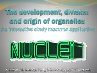
The development, division and origin of organelles. An interactive study resource application. A. Dhillon, M. Farfan, I. Perry, S. Sanchez-Leon, M. Watkinson. How the program works. Navigate using: The next arrow. The previous arrow. The home button. The topic jump button.

E N D
The development, division and origin of organelles An interactive study resource application A. Dhillon, M. Farfan, I. Perry, S. Sanchez-Leon, M. Watkinson.
How the program works Navigate using: The next arrow. The previous arrow. The home button. The topic jump button. See more using: The extra info button. The Movie button. The Audio button.
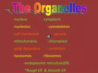
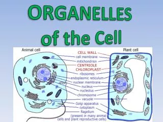
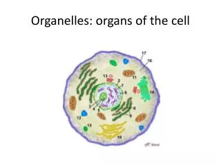
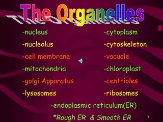
Topic index The Origin of organelles The Division of organelles The Development of organelles Review Quiz References
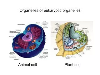
Organelle origin Autogenously Origin theory: Example of autogenously origin of the flagellum Here it is thought that actin and micro tubules converging around the nucleus forced a protrusion of the cell in a particular direction down to targeted traffic of cellular membrane components. This developed into a conserved structure and through small modifications gained order to the structure of the organelle. Gradual development and modification of proteins saw a mechanical turning force created. The cytomembrane system: It wasmost likely originated from invaginations and infoldings of the plasma membrane of some wall-less ancestral cell. The cell size increased and then all these infoldings and invaginations grew deeper and more convoluted, splitting into vesicles. This entire network gradually differentiated into more specialized parts, as the rough and the smooth endoplasmic reticulum (ER), the nuclear envelope, the Golgi complex, endosomes and lysosomes. An evidence of this is the similarities between the co-translational protein translocation system in bacteria membranes and in rough ER. The nucleus: The eukaryotic nucleus is surrounded by an envelope; which is typically made of rough ER vesicles derived from the invagination of the plasma membrane, and cytoskeletal elements (lamina) that are associated with each other and with pore complexes. In the process of mitosis this complex assemblage dissociates and reassembles into distinct envelopes that surround the two sets of daughter chromosomes. The formation of the nuclear envelope must have accompanied or followed the development of the cytomembrane and cytoskeletal systems. Prokaryotic chromosomes are anchored to the cell membrane. Perhaps a vesicle derived from the piece of membrane that bore the chromosome joined with other vesicles to form a double-membranous envelope around the genetic material. So now we have a structure which allows the physical separation of DNA replication and transcription from RNA translation, which occurs in the cytoplasm. This fact seems to act as the evolutionary driving force for their development so it is likely that the formation of the nuclear envelope initiated the entire succession of events that culminated into the eukaryotic nucleus with linear chromosomes and their organization with histones into nucleosomes and chromatin fibers, Nucleoli, spliceosomes, traffic-regulating pore complexes and a new model of division, mitosis. close Figure: 1 Flagellum autogenously origin theory 1 The nucleus
Organelle Origin Endosymbiotic Origin theory: There are two classes of organelles that are surrounded by two membranes: plastids and mitochondria. Both of these organelles are of endosymbiosis origin as it defended the Endosymbiosis Theory, reformulated by Lynn Margulis2. These organelles have a different biogenetic pathway. There are some evidences supporting the endosymbiosis theory which suggest that a bacterial endosymbiont established itself inside on a protoeukaryote, or what it the same, an originally free-living prokaryotic organism was engulfed by a “eukaryotic cell”. Some of these evidences are: -No nucleus, and own circular genome with lack of histone proteins in plastids and mitochondria. -They synthesize their own proteins. -Prokaryotic-like ribosomes, polymerases and enzymes. -Small size and shape. -Type of division which is encoded by several homologues of prokaryotic genes that are in the nucleus. - Different composition of the outer and inner membrane. Figure: 2 Temporal positioning and phase progression of Mitochondrial and Plastid Endosymbiosis3 Play movie Exit movie
Organelle Origin Endosymbiotic Origin theory: Eukaryotic cells most probably acquired mitochondria and plastids after they had developed the cytomembrane and cytoskeletal machineries that are involved in the endocytic uptake of extracellular materials, and not before, so it is possible to the cell engulf the symbiont. Then a mutual benefit relationship is established between both organisms, it also permit a compartmentalization of function 4 5. Phylogenetic analysis suggests that Mitochondria developed from an alpha-proteobacteria and chloroplasts from cyanobacteria that were taken inside of another cell as endosymbiont, taking mutual benefits. The acquisition of both organelles were independent events, mitochondrion endosymbiosis probably preceded the plastids one. Then complex molecular event drove the evolution of this endosymbionts into contemporary semi-autonomous organelles. A critical step in the transition from autonomous endosymbiont to organelle was the sharing of genetic information with genes transfer to the nucleus. Also many of endosymbiont genes have been lost (genome reduction), and most of the retained ones were transferred to the nucleus. Figure: 3 Stages in Endosymbiosis
Organelle Division • Chloroplast Division: • The first step in chloroplast division sees a ring forming around the plastid on the inside and outside with Fts Z1 & 2 proteins. To this ARC 6 protein binds and co-ordinates the structural formation of the ring. Another protein, Min D is found at either end of the plastid and helps in centralising the ring. • ARC 5 is a “Dynamin like” protein and works to constrict the newly formed PD ring. Rather than depolymerizing the ring to pull as in microtubules the ring instead coils tighter pulling the cell membrane to a pinch called the isthmus where the membrane breaks to form two new plastids 7 • Mitochondrial Division & Fusion: • Mitochondria divide by Binary fission (known as mitochondrial fission), similar to chloroplasts. • The Process carried by: • Division ring Proteins - Drp1 & • Mitochondrial fission 1 proteins – Fis1 • Step 1. Drp1 is orchestrated by Fis1, which is found on the outer mitochondrial membrane. Fis1 attracts Drp1 to mitochondrial fission sites. • Step 2. Drp1oligomers combine in a curved formation around the outer surface of the mitochondria. • Step 3. The curved Drp1 structures then constrict, this pinches off the mitochondria two produce two copies of the organelle. Constriction occurs through the utilisation of energy from GTP hydrolysis 8 9. • It has also been observed that mitochondria can fuse. This sees a set of proteins on each mitochondria called Mfn1 set which join in a region called the HR2. These coiled alpha helices pull the two mitochondria together until the mitochondria membranes can fuse 10 Figure: 4 Chloroplast division 7 Figure: 5 Mitochondrial division 11
Organelle Division Factors affecting Plastid Division in Higher Plants Intro Nutrition Light Diurnal Temperature
Organelle Development The Peroxisome A peroxisome is a single membrane structure containing digestive enzymes in crystalline form. They are used in the catabolism of long chain fatty acids and other complex lipids like amino acids. They work through the pentose phosphate pathway and account for up to 10% of a cells activity. There are two current models for its development in a cell. The first, growth and division, sees Px proteins and other membrane bound proteins being incorporated together. The second, maturation model, sees Px vesicles that have derived from the endoplasmic reticulum in different locations come together and fuse before maturing. More recent evidence suggest the need of ATP in the recycling of its receptors 13 14 . Figure: 6 The different models of Peroxisome development and maturation 12
Organelle Development • Lysosome development • Lysosomes are membrane bound organelles found in animal and plant cells. They vary in shape, size and number per cell and appear to operate with slight differences in cells of yeast, higher plants and mammals. Lysosomes contain many different enzymes and contribute to recycling substances as they assist with degrading material taken in from outside the cell and expired components from within the cell. Lysosomes don’t “choose” which cells are ingested/recycled. This is a function of the processes of programmed cell death (apoptosis) and phagocytosis. In humans, errors in the genetic code account for about 30 lysosomal storage disorders. Lysosomal storage diseases can be caused by: • Enzymes present within the lysosome that don’t function properly • Enzymes that are not transported into the lysosome • Enzymes that are not produced • In both the maturation and vesicular transport models late endosomes develop to become a lysosome. • In the maturation model an early endosome is formed from vesicles originating in the plasma membrane combining together. Various other vesicles deliver and remove chemicals until the late endosome, and then the lysosome stage is reached. • In the vesicle transport model, early and late endosomes are considered stable separate organelles with vesicles carrying chemicals from early endosomes to late endosomes. Vesicle transport from the golgi to the lysosome requires microfibrils, Late endosomes then mature to become lysosomes 17. Figure: 7 Lysosome development 15
Organelle review True or False Click on correct answer T F Q1: Mitochondria use fusion and fission to control shape and function T F Q2: Mitochondria are thought to have evolved from Cyanobacteria T F Q3: Mitochondria fuse via the HR3 regions F T Q4: microtubules coil tighter to pinch the plastid in chloroplast division
Organelle review Gap fill Work out which words go in which gap. When you think you are ready, click reveal to see if your right. Chloroplasts are ………….. organelles, and are part of the endosymbiosis theory. This is where an originally free ………….organism is engulfed by a eukaryotic cell, they then form a symbiotic relationship in which they share energetic ……….. Chloroplasts are said to have evolved from ………… due to their photosynthetic properties. All chloroplasts are derived from …………., many other plastids also derive from this common source, and these include: When light is present the inner membrane of the proplastid will bud and produce vesicles which will then form thylakoid vesicles and finally stroma and……., giving a mature chloroplast (…-…µm). Semi-autonomous, Protobacteria, plastids, autonomous, amyloplasts, 20-30µm, Prokaryotic, Cyanobacteria, fungi, Grana, 40-50µm, amoeba, 10-20µm, products Reveal Chloroplasts are semi-autonomous organelles, and are part of the endosymbiosis theory. This is where an originally free prokaryotic organism is engulfed by an eukaryotic cell, they then form a symbiotic relationship in which they share energetic products. Chloroplasts are said to have evolved from cyanobacteria due to their photosynthetic properties. All chloroplasts are derived from proplastids, many other plastids also derive from this common source, and these include: When light is present the inner membrane of the proplastid will bud and produce vesicles which will then form thylakoid vesicles and finally stroma and grana, giving a mature chloroplast (10-20µm).
Organelle review Process order Try order the process of chloroplast division. When you think you are ready, click reveal to see if your right. • 1. A dynamin ring is formed • 2. The FtsZ ring forms • 3. Dynamin-like proteins associates • 4. The chloroplasts separate and the remnants of the PD and dynamin ring remain. • 5. The PD ring forms and associates with the outer and inner envelope membranes • 6. The ring constricts forming an isthmus Reveal Correct order: 2, 5, 3, 1, 6, 4
Organelle review Match up: predict what the answer will be. Click on the correct answer From which organelle are the vesicles derived from in the peroxisome maturation model? Nucleus Golgi apparatus Wrong Endoplasmic reticulum Lysosomes What’s the name of the pathway in which newly synthesised peroxisome proteins are incorporated into pre existing /recently divided peroxisomes? Growth and division model Endosymbiotic model Wrong Maturation model Recombinant theory
References • Alberts, Johnson B., Lewis A.J. (2002) Molecular Biology of the Cell. 4thediton, New York, Garland Science • Dyall S.D., Brown M.T., and Johnson P.J. (2004) Ancient Invasions: from Endosymbionts to Organelles. Science 304, 253-255. • Becker W., Kneinsmith L., Hardin J., and Bertoni G. (2000) The World of the Cell. Seventh edition, 86. 298-9 • Duve C.D. 2007. The Origin of eukaryotes: a reappraisal. Nature Reviews, 8, 395-403. • Martin W. (2010) Evolutionary origins of metabolic compartmentalization in eukaryotes. The Royal Society, 365, 847-852. • Bhattacharya D., Archibald J.M., Weber A.P.M., and Reyes-Prieto A. (2007) How do endosymbionts become organelles? Understanding early events in plastid evolution. BioEssays, 29, 1239–1244. • Miyagishima S (2009) Dividing the photosynthetic spoils. Research Highlights: Biology 4. • Giezen M.V.D.(2011) Mitochondria and the Rise of Eukaryotes. BioScience, 61, 594-599. • Hales K. (2010) Mitochondrial Fusion and Division. Nature Education, 3(9):12. • Baloh R.H., Schmidt R.E., Pestronk A., and Milbrandt J. (2007) Altered axonal mitochondria transport in the pathogenesis of Charcot-Marie-Tooth disease from Mitofusin 2 Mutations. The Journal of Neuroscience, 27:2: 422-430 • Youle R, and Karbowski M (2005) Mitochondrial fission in apoptosis. Nature reviews: Molecular cell biology 1697: 622 - 634. • Klei I, and Veenhuis M (2002) Peroxisomes: flexible and dynamic organelles. Current opionion in Cell Biology: pp. 500-505. • Latruffe N, and Vamecq J (2000) Evolutionary aspects of peroxisomes as cell organelles, and of genes encoding peroxisomal proteins. Biology of the Cell, 92:6, 389-395 • Lazarow L.P. (2003) Peroxisome biogenesis: advances and conundrums. Current Opinion in Cell Biology, 15:489-497 • Luzio J.P., Rous B.A., Bright N.A., Pryor P.R., Mullockand B.M., and Piper R.C. (2000) Lysosome-endosome fusion and lysosome biogenesis. Journal of Cell Science, 113, 1515-1524 • Chen H., Vermulst M., Wang Y.E., Chomyn A., Prolla T.A., McCaffery J.M., et al. Mitochondrial Fusion Is Required for mtDNA Stability in Skeletal Muscle and Tolerance of mtDNA Mutations. Cell. 2010 16 April 2010;141(2):280-9 • Pramanik S., Nerad J. (2007). Chronic Progressive External Opthalmoplegia (CPEO)- Kearns-Sayre Syndrome: 12 y.o boy with painless, progressive ptosis OU over 3 years. Available: http://webeye.ophth.uiowa.edu/eyeforum/cases/case24.htm. Last accessed 15/11/2011 • Music from Melodysheep“We are all connected”
Contact us Join our Facebookpage for free download! http://www.facebook.com/#!/groups/194154237334594/
Organelle Division Mitochondrial Fusion: What Happens when it Goes wrong? Mitochondria require fusion in order to remain stable. They fuse together to mix up their contents and their mtDNA. When fusion is blocked, mitochondria will become smaller and smaller in size when they divide. The smaller mitochondria will be unfit to produce the energy requirement of a cell. They also accumulate more "point mutations and deletions“ which are harmful to them. The researchers hope that studies conducted on rodent mitochondria will one day help to provide a cure for mitochondrial encephalomyopathies that affect humans 16. Figure: 8The effects of blocking fusion in rodent mtDNA . We can see a reduction in size as well as an increase in mutations.16 Mitochondrial diseases associated with division include: Parkinson’s Alzheimers Kearns-sayre syndrome Pearson’s syndrome Leigh’s syndrome
Organelle Division Mitochondrial Fusion: What happens when it goes wrong? • Kearns-Sayre Syndrome • This is a rare mitochondrial disorder, and is characterised by the onset of opthalmoparesis (paralysis of one or more extraocularmuscles) and pigmentary retinopathy before 20 years old. Other frequently associated clinical features include: • Cerebellar ataxia • Cardiac conduction block • Raised cerebrospinal fluid protein content • Affected children have short stature and often have multiple endocrinopathies • The mitochondrial genome is a closed circular loop of double stranded DNA found in multiple copies within the mitochondrial matrix. Kearns Sayre Syndrome occurs as a result of large-scale single deletions/rearrangements of mitochondrial DNA, which are usually not inherited but occur spontaneously, probably at the germ-cell level or very early in embryonic development. The risk of maternal transmission has been estimated to be approximately 1 in 24. The deletions vary in size and location on the mitochondrial genome in different individuals. Figure: 9 Kearns-Sayres Phenotype progression 17
Organelle Division PeroxisomeDevelopment: What happens when it goes wrong? Zellweger syndrome This is one of four of the diseases called peroxisomes biogenesis disorders (PBD). These are inherited conditions that damage the white matter of the brain and also affect how the body metabolises particular substances in the blood and organ tissues. The diseases are caused by defects in any 1 of 13 genes, termed PEX genes (these are required for normal function of the peroxisome). Peroxisomes are required for normal brain development and function and the formation of myelin, the whitish substance that coats nerve fibers. In this disorder the peroxisome fails to develop with the correct lipid composition from the endoplasmic reticulum. Symptoms of these disorders include an enlarged liver, characteristic facial features and neurological abnormalities. There’s no cure for Zellweger syndrome or course of treatment. Prognosis is poor for infants and many don’t survive past the first 6 months (geneva foundation for medical education). Figure: 10 The effects of incorrect peroxisome development on an infant. (Geneva foundation for medical education)