Download
1 / 42
440 likes | 509 Vues
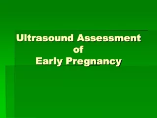
This informative text guides you through the ultrasound assessment of early pregnancy stages, from ovulation to organogenesis, highlighting key events like fertilization, implantation, and the development of germ layers. Learn about the establishment of vital structures and the evolution from embryo to fetus, including organ formation and circulatory system development. Helpful for medical professionals and expectant parents alike.

E N D
from OVULATION THE ONLY PERIOD OF GESTATION NOT DETECTED DIRECTLY IMPLANTATION to
First Week of DevelopmentOvulation to Implantation(not visible by ultrasound) Fertilization occurs at the ampullary region of the fallopian tube. The diploid number of chromosomes are restored. Chromosomal sex is determined. In the fifth day, the blastocyst is embedded in a well prepared, thick endometrium.
UTERINE ARTERY ARCUATE ARTERIES RADIAL ARTERIES CURVED BRANCHES (spiral art.) ZONE FUNCTIONALIS LINEAR BRANCHES (basal art.) ZONE BASALIS
SECRETION PHASETHICKNESS 7 - 16 mm • homogen and hyperechogenic echo as result of mucin and glycogen in tortuotic endometrial glands
Blastocyst 5 days post conception
Second week of Developmentbilaminar Germ Disk(not visible by ultrasound) • The trophoblast differentiates into an inner cell mass (the cytotrophoblast) and an outer cell mass (the syncytiotrophoblast), which erodes the endometrium. • Lacunar network is formed by the end of the second week and a primitive utroplacental circulation begins.
Third week of Developmenttrilaminar germ disk(not visible by ultrasound) • The most characteristic event is gastrulation. • By the end of the third week three basic germ layers consisting of ectoderm, mesoderm, and endoderm are established. • Tissue and organ differentiation has begun.
Third to Eighth Week of DevelopmentThe Embryonic Period • This is the period of organogenesis. • Each of the three germ layers (ectoderm, mesoderm and endoderm) give rise to its own tissues and organ systems. • Major features of body form are established.
Establishment of intervillous circulation • Lacunar formation – 10th to 13th days after conception • Filled with blood on day 15th • Tertiary Villi formation on day 20th Villous capillaries become connected with the embryonic heart tube
THE ROLE OF YOLK SAC - TRANSFER OF NUTRIENTS IN THE 3rd AND 4th WEEK OF GESTATION - HAEMATOPOESIS IN THE 5TH WEEK - THE INITIAL SITE OF PRODUCTION OF AFP, PREALBUMIN, ALBUMIN AND TRANSFERIN - ALL FUNCTIONS ARE COMPLETED BY 8 WEEKS OF GESTATION GESTATIONAL SAC DIAMETER › 8 mm
5-6 weeks: • Early trophoblast • Lacunar flow • Secondary yolk sac • the first visible • structures within • gestational sac
INTERVILOUS SPACE ( IVS ) 5 - 6 w.g.a.
Embryo at 6 wks The embryo is 3-4 mm. G.S is 14-15 mm Heart activity visualized
6 WEEKS EMBRYO INTERVILLOUS BLOOD FLOW ONSET OF HEART ACTIVITY
7 WEEKS LIMB BUDS GROSS BODY MOVEMENTS FETAL AORTA UMBILICAL CORD FETAL HEART THE HEAD IS MORE PROMINENT DUE TO THE DEVELOPING RHOMBENCEPHALON. NO EVIDENCE OF CEREBRAL CIRCULATION.
Embryo at 7 wks Chorionic cavity 19-20 mm Embryo 5-6 mm
8 WKS Cerebral circulation started CEREBRAL VESSELS FETAL AORTA GROSS BODY MOVEMENTS ARMS AND LEGS MOVEMENTS
Embryo at 8 wks Amniotic Cavity 17-19 mm. Embryo size is 16-18 mm. Choroinic cavity 30-32 mm.
Third month to birth the Fetus and Placenta • The fetal period extends from the ninth week of gestation until birth and it is characterized by rapid growth of the body and maturation of organ systems.
Cerebral circulation is established 9 WEEKS CEREBRAL VESSELS HEART FETAL AORTA
9 WEEKS STARTLE GROSS BODY MOVEMENTS STRECTHING ARMS AND LEGS MOVEMENTS HEAD ROTATION HAND MOVEMENTS
Fetus at 9 wks with clear amniotic membrane Embryo is 24-31 mm
Fetus at 10 wks Beginning of Ossification Falx cerebri appears Choroid plexus occupy the ventricles Abdomen shows physiological omphalocele
3D Fetus at 10 wks ALL THREE SEGMENTS OF THE UPPER AND LOWER EXTREMITIES ARE VISIBLE
Fetus at 10 wks showing facial detail The neck is visualized which in a sagital section presents the nuckal area – a double hyper echogenic outline – septated by a millimetric hypo echogenic band corresponding to subcut. tissue.
11 WEEKS • GENERAL MOVEMENTS • STARTLE • STRETCHING • ISOLATED ARM MOVEMENTS • ISOLATED LEG MOVEMENTS • HEAD MOVEMENTS AND ROTATIONS • HAND TO FACE CONTACT
12 weeks Head rotation Limb movements General movements Clench and unclench fists
Fetus at 12 wks – arm/fingers visible The fetus is much more explorable
13 weeks • GENERAL MOVEMENTS • STARTLE • STRETCHING • ISOLATED ARM MOVEMENTS • ISOLATED LEG MOVEMENTS • HEAD MOVEMENTS AND ROTATIONS • HAND-FACE CONTACTS
BICHORIONIC BIAMNIOTIC TWINS LAMBDA SIGN 3D
3D TRIPLETS FRONT BACK
3D QUINTUPLET QUADRIPLET
2D SONOEMBRYOLOGY GESTATIONAL WEEKS
THANK YOU