Download
1 / 16
300 likes | 944 Vues
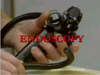
Dr. Alaa Hussien Ali AL-NASER. Endoscopy in gynecology. Introduction:

E N D
Dr. Alaa Hussien Ali AL-NASER Endoscopy in gynecology
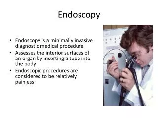
Introduction: The recent development of sophisticated optic systems providing broader viewing angles, finer resolution and magnification, and significantly improved fiberoptic lighting sources has rapidly advanced the endoscopic aspects of gynecologic diagnosis. The endoscopic investigations include hysteroscopy for the magnified viewing of the uterine cavity, culdoscopyandlaparoscopyfor the assessment of intrapelvic organ pathology and function.
1- Hysteroscopy: Diagnostic accuracy and enhanced therapeutic capabilities are achievable with the availability of hysteroscopy for the patient with symptoms pointing to intrauterine pathological conditions. The procedure: The hysteroscope is a telescope with an outer dimension of up to 8 mm, which is inserted transcervically after dilatation of the cervix, under paracervical anesthesia or G.A. The uterine cavity can be distended with 5% dextrose in water, normal saline, 1.5% glycine, 32 % dextran solution, or carbon dioxide gas insufflation.
The indications for diagnostic hysteroscopy 1- Evaluation of abnormal bleeding in the reproductive age or postmenopausal women. 2- Assessment of infertility status. 3-Localization and removal of intrauterine foreign bodies (IUDs or plastic instruments). 4- Evaluation of congenital uterine anomalies or adhesions. 5-Localization and excision of endometrial polyps or submucous leiomyomata.
The contraindications to the procedure • 1-Active infection. 2-Acute hemorrhage. 3- Known pregnancy. • Intrauterine visualization by hysteroscopy has added a significant dimension to the management of patients with relatively common gynecologic problems. The technique increases the accuracy of diagnosis and allows relatively simple therapy in some cases. In particular, the localization and removal of IUDs through the hysteroscope has provided a much needed alternative to the use of x-ray films of the pelvic area and blind intrauterine manipulations for retrieval of devices.
Operative hysteroscopy,often performed under laparoscopic control, has been widely used to incise or remove congenital uterine septa, sumucous myomas, or adhesions for transcervical sterilization and for the ablation of the endometrium in medically compromised patient with refractory menorrhagia.
Complications: • Anesthetic complications • Perforation of the uterus • Bleeding due to myometrial vascular traum • Thermal trauma to adjacent structure • Complication related to distension media: 1- CO2 embolization 2-Glyceine fluid overload& electrolyte imbalance (fetal)
2- Laparoscopy: The procedure : The patient is anesthetized and placed in a modified lithotomy position, and the bladder is emptied. A Rubin’s cannula is inserted into the cervix & uterus and thereby enhance visualization. It also permits the injection of dye to observe tubal patency. Intubation anesthesia is generally advocated because of the steep Trendelenburg position required for upward movement of the intestine and because of the large amount of carbon dioxide that exerts diaphragmatic pressure.
2- Laparoscopy (cont.) A needle is inserted just below the umbilicus in the midline, and carbon dioxide is instilled under controlled pressure to a volume of 3 liters.
2- Laparoscopy (cont.) • After abdominal distention, the needle is withdrawn. The small intraumbilical incision is enlarged and a trocar with its sheath is introduced through the same opening.
2- Laparoscopy (cont.) • Continuous gas insufflation under controlled pressure maintains abdominal distention. The trocar is then removed and replaced by the laparoscope. Fiberoptic light sources are attached to the laparoscope and pelvic visualization is begun. It is often both helpful and necessary to introduce a blunt probe through a separate small trocar incision to assist in manipulation of the pelvic organs for complete examination.
Indications for laparoscopy: A. diagnostic 1- infertility primary , secondary assess the following a. POD b. tubes(adhesion,pyosalpinx,patency) c. ovaries(size,shape, pathlogy like cyst) d. Curettage done at some times to look for any TB. Granuloma and check ovulation. 2- amenorrhea(1,2): Pcos,absent ovary, infantile uterus, menopause. 3- chronic pelvic pain to diagnosed PID and endometriosis. endometriosis, endometrial adhesion. 4- lower abdominal pain, acute pelvic pain such as acute salpingitis, ectopic pregnancy, torsion of the ovary and ovarian cyst. 5- nature of any pelvic mass(infected, cystic, solid) 6- diagnosis trauma of uterus during curettage. 7- f0llow up of malignancy or tumor in general. Theraputic indication 1- excision or evaporation of endometriosis 2- relieve of any adhesion 3- tubal sterlization,surgury, salpingectomy, or salpingestomy. 4- ovarian cyst excision, oophorectomy, ovarian diathermy in pcos. 5- bowel adhesion removal 6- myomectomy or myolysis 7- repair of uterine perforation 8- laparoscopic hysterectomy 9- ventro suspension of uterus 10- oocyte retrieval in IVF 11- removal of IUCD
Complication of laproscopy: • A- Hg. Occure in vesseles such as aorta, inferior vena cava, iliac vesseles by needle or canula so we perform syringe test. • B- air embolism during peritoneal insufflation. • C- surgical emphysema that occure if needle pass through subcutaneous tissue then insufflation. • D- viseral damage s.a bladder, bowel specially in previous surgury or adhesion. • E- cardiopulmonary accident(excessive insuffulation lead to pr. On diaphragm and IVC lead to increase cardiac out put so cardiac shock. • F- failure of procedure
Contraindication of laparoscopy: • Obsolute contraindication • 1- mechanical and praltic ileus. • 2- large abdominal mass • 3- generalised peritonitis Relative contraindicat. • 4- irreducible hernia 1. multiple incision • 5- recent MI 2. sepsis • 6- cardiac failure 3. IHD • 7- cardiac conductive defect 4. coagulopathy • 8- respiratory failure 5. hiatus hernia • 9- sock 6. gross obesity • 10- sever restrictive air way disease.
culdoscopy Is a technique for visualizing the pelvic organs under local anesthesia with the patient in the knee-chest position through a puncture of the cul-de-sac. Culdoscopy has generally been replaced by laparoscopy because of difficult orientation, limited field of vision and higher failure rate. Nevertheless, it remains a useful endoscopic accessory for diagnosis of gynecologic pelvic pathology and obviates the need for a general anesthesia.