Download
1 / 50
560 likes | 1.02k Vues
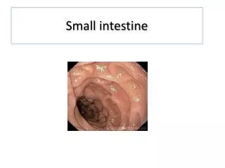
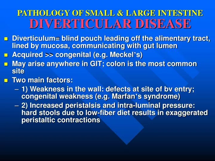
PATHOLOGY OF SMALL & LARGE INTESTINE DIVERTICULAR DISEASE. Diverticulum= blind pouch leading off the alimentary tract, lined by mucosa, communicating with gut lumen Acquired >> congenital (e.g. Meckel ’ s) May arise anywhere in GIT; colon is the most common site Two main factors:

E N D
PATHOLOGY OF SMALL & LARGE INTESTINEDIVERTICULAR DISEASE • Diverticulum= blind pouch leading off the alimentary tract, lined by mucosa, communicating with gut lumen • Acquired >> congenital (e.g. Meckel’s) • May arise anywhere in GIT; colon is the most common site • Two main factors: • 1) Weakness in the wall: defects at site of bv entry; congenital weakness (e.g. Marfan’s syndrome) • 2) Increased peristalsis and intra-luminal pressure: hard stools due to low-fiber diet results in exaggerated peristaltic contractions
PATHOLOGY OF COLONIC DIVERTICULOSIS • Colonic diverticulosis is common in the West (50% of adults >60 yrs); associated with low-fiber diet • Multiple small 0.5-1 cm • Sigmoid colon is involved in 95% of patients • Gross examination: Prominent taeniae coli & circular muscle bundles due to muscular hypertrophy; outpouching may be visible from serosal surface • Histology: sacs with thin wall made of mucosa & submucosa surrounded by fat or intact peritoneum
CLINICAL FEATURES OFCOLONIC DIVERTICULOSIS • Usually asymptomatic • Intermittent cramping or continuous LLQ discomfort, sensation of incomplete emptying of rectum • Complications: • Diverticulitis: accentuates symptoms, tenderness, fever • Peridiverticulitis, perforation, abscess or fistula • Fistula formation • Bleeding • Rx: High-fiber diet; surgery
PATHOLOGY OF SMALL & LARGE INTESTINEBOWEL OBSTRUCTION • May occur at any level, but is more frequent in small intestine • May be acute or chronic • Clinical features: abdominal distension (+/- pain) , followed by vomiting, then constipation • May present at birth, during infancy, childhood or adulthood • Obstruction may be in the lumen, wall or extramural • Tumors and infarction account for 10-15% of small bowel obstruction • Hernias, intestinal adhesions, intussusception, and volvulus account for 80% of cases
CLASSIFICATION OFBOWEL OBSTRUCTION • Mechanical: • Congenital: strictures, atresias, bands, imperforate anus, meconium in cystic fibrosis • Infants: hernias, intussusception • Adults: hernias, adhesions, tumors, inflammatory strictures, obstructive gallstones, fecaliths, foreign bodies, volvulus • Functional: • Paralytic ileus: electrolyte imbalance, neural injury, or reflex atony secondary to peritonitis • bowel infarction, • myopathies & neuropathathies (e.g. Hirschsprung’s)
BOWEL OBSTRUCTIONHERNIAS • Weakness or defect in wall of peritoneal cavity with protrusion of a serosa-lined sac of peritoneum • Inguinal & femoral canals, umbilicus & surgical scars • Segments of viscera may protrude & become entrapped, mostly in inguinal hernias • If small bowel is involved, partial or complete obstruction of its lumen may follow • Incarceration: permenant trapping of hernial sac contents due to venous stasis & edema • Strangulation: Venous & arterial supply to entrapped viscus is compromised leading to infarction or gangrene
BOWEL OBSTRUCTIONINTESTINAL ADHESIONS • Due to localized or generalized peritonitis secondary to: • Surgical procedures • Infection • Endometriosis • Rarely due to congenital fibrous bands • May result in intestinal obstruction, incarceration and strangulation • Fibrous bridges which develop between bowel segments creating closed loops through which viscera may slide & become entrapped (internal hernias)
BOWEL OBSTRUCTIONINTUSSESCEPTION • Uncommon disorder where a segment of small intestine, constricted by a a wave of peristalsis, suddenly becomes telescoped into the immediately distal segment of bowel • Intussusceptum: trapped bowel segment • Intussuscipiens: bowel segment which envelops it • Mostly in infants & children of unknown pathogenesis • In adults, an intraluminal tumor may act as point of traction, pulling along a segment of bowel • Intestinal obstruction • Intestinal infarction due to trapping of mesenteric blood vessels
BOWEL OBSTRUCTIONVOLVULUS • Complete twisting of a loop of bowel about its mesenteric base of attachment • Mostly in small intestine; large intestine especially sigmoid & cecum, and other structures (e.g. ovary) may be involved • Constriction of venous outflow &/or arterial supply • Intestinal obstruction and infarction • Uncommon disorder, occuring in all ages when an intestinal segment becomes longer & the mesentary narrower • May be caused by colon malrotation & peritoneal bands
SMALL & LARGE INTESTINETUMORS • Colonic tumors are much more common than small intestinal tumors • May arise from any layer of GIT wall (mucosa, submucosa, muscularis or serosa); most are of epithelial origin • The majority are benign; however, colonic cancers are a major cause of morbidity & mortality • Clinical presentation: • Asymptomatic; incidental finding • Blood per rectum; anemia • Bowel obstruction
INTESTINALTUMORS • Benign: • Stromal tumors • Hyperplastic & adenomatous polyps • Lipomas • Malignant: • Adenocarcinoma • Carcinoid tumors • Lymphoma • Sarcoma
PATHOLOGY OF SMALL & LARGE INTESTINECARCINOID TUMORS • Tumors of neuroendocrine cells, which are widely distributed cells of epithelial stem cell origin capable of producing a variety of bioactive compounds • Tumors may produce multiple compounds or a predominant product, causing a clinical syndrome such as gastrinoma, insulinoma ... • Tumors arrise in GIT, lung, biliary tree & pancreas • 50% of small intestinal & 2% of colonic tumors • Any age, peak incidence in 6th decade • Carcinoids are well differentiated tumors that are potentially malignant tumors and may metastasize
CLINICAL FEATURES OFCARCINOID TUMORS • Sites: appendix>small intestine (ileum)>rectum> stomach>colon • Most are asymptomatic (particularly appendix) • May cause local symptoms (angulation or obstrcution of small intestine) • Secretory products may produce a variety of endocrinopathies, e.g. Zollinger-Ellison syndrome, Cushing’s syndrome, hyperinsulinism • Carcinoid syndrome: 1% of patients & 20% of those with widespread metastasis; due to serotonin secretion; vasomotor disturbances, intestinal hypermotility, hepatomegaly and systemic fibrosis
PATHOLOGY OF CARCINOID TUMORS • Gross: usually small tumors, rarely >3 cm, forming an intramural or submucosal yellow-tan firm elevation or polypoid lesion • Histology: monomorphous population of cells with scant granular cytoplasm & round nuclei. Neoplastic cells form islands, nests, strands or glands. Intact or ulcerated overlying mucosa. • May be multicentric (stomach, ileum) • May be localized or have local spread &/or metastasis to lymph nodes or more distant sites, particularly liver
CLINICAL BEHAVIOUR OFCARCINOID TUMORS • Clinical behaviour is difficult to predict from histologic features • Behaviour but generally depends on: • 1) Size: >2 cm and those invading >1/2 wall thickness • 2) Site: appendiceal & rectal carcinoids almost never metastasize • 5 years survival rate is excellent: 90% • 5 years survival rate for small intestinal tumors with liver metastasis: 50% • Widespread disease usually causes death
SMALL INTESTINALADENOCARCINOMA • Most arise in duodenum, including ampulla of Vater • Polypoid fungating tumor growing in a napkin-ring encircling pattern • Early on, tumor is asymptomatic • Signs & symptoms usually appear late, when tumor has already penetrated bowel wall into mesentery, other bowel segments or spread to LN, or metastasize to liver or more widely • c/o symptoms of abdominal obstruction (pain, nausea, vomiting, and weight loss) • 5 yrs survival with wide en bloc excision: 70%
SMALL & LARGE INTESTINEPOLYPS • Polyp= a tumorous mass protruding into the lumen of the gut; may be pedunculated or sessile • Major types: • Non-neoplastic polyps: about 90% of polyps; formed as a result of abnormal mucosal maturation, inflammation or architecture • Hyperplastic polyps • Hamartomatous polyps • Inflammatory polyps • Neoplastic polyps: formed as a result of epithelial proliferation and dysplasia, i.e. pre-cancerous • Adenomatous polyps
INTESTINAL POLYPSHYPERPLASTIC POLYPS • Majority of polyps • Site: mostly in colon, >50% in rectosigmoid • Patients: increase in frequency with age • Appearance: Most are small <5 mm, nipple-like protrusion; multiple or single • Pathology: histologically consist of abundant crypts lined by well-differentiated goblet or absorptive epithelial cells, seperated by scant lamina propria • Asymptomatic or blood &/or mucus per rectum • No malignant potential
INTESTINAL POLYPSHAMARTOMATOUS POLYPS • Juvenile polyps: • Hamartomatous proliferation of lamina propria surrounding cystically dilated glands • Patients: usually <5 yrs; in adults called “retention polyp” • Appearance:1-3 cm in children; smaller in adults • Single rounded or lobulated, +/- stalk • Bleeding per rectum; stalk may twist and undergo painful infarction • No malignant potential
INTESTINAL POLYPSHAMARTOMATOUS POLYPS • Peutz-Jeghers polyps: • Rare autosomal dominant syndrome, characterized by GIT polyps, melanotic mucosal and cutaneous pigmentation around lips, oral mucosa, face, genitalia & palmar surfaces of hands • Site: polyps may occur in stomach (25%), colon (30%) and small intestine (100%) • Appearance: large peduculated lobulated polyps • Pathology: branching framework of connective tissue & smooth muscle • Polyps have no malignant potential, but PJS patients have increased risk of developing carcinoma of pancreas, breast, lung, ovary & uterus
INTESTINAL POLYPSADENOMATOUS POLYPS • Vast majority of adenomas arise in colon • Variable in size; pedunculated or sessile • Results from epithelial proliferation and dysplasia, which may range from mild to carcinoma in situ • Three architectural types: • 1) Tubular adenoma • 2) Villous adenoma • 3) Tubulovillous adenoma • Risk of malignant transformation depends on: • Polyp size: <1 cm (v. rare); 1-2 cm (10%); >2 cm (45%) • Histologic architecture: proportion of villous component • Severity of dysplasia
INTESTINAL POLYPSADENOMATOUS POLYPS • Tubulovillous adenoma: 5-10% of adenomatous polyps; mixture of tubular & villous (20-50%) areas; intermediate in frequency of having a stalk, size, degree of dysplasia & risk of carcinoma • Clinical features: 20-30% are <40 yrs; 50% after 60 • Small polyps: asymptomatic • Occult bleeding, anemia, hypoproteinemia & hypokalemia • Intestinal obstruction or biliary obstruction • Rx: Most are cured by adequate endoscopic excision • Px:Annual endoscopic follow-up
>85% of adenomas Majority in distal colon; 50% in rectosigmoid Most are small (few mm-2 cm) with a stalk (-2 cm) Histology shows neoplastic epithelium forming glands & tubules Variable degrees of dysplasia to CIS Invasive carcinoma rare 1% of adenomas Rectum & rectosigmoid 75%, followed by cecum & ascending colon Sessile ranging from 1-10 cm (most are 1-3 cm) Histology shows neoplastic cells forming villi (>50% villous) Carcinoma in situ 10% Invasive carcinoma 30% ADENOMATOUS POLYPSTUBULAR ADENOMA VILLOUS ADENOMA
INTESTINAL POLYPSFAMILIAL POLYPOSIS SYNDROMES • Familial adenomatous polyposis (FAP): • Rare autosomal dominant trait • Dx: at least 100 polyps (average 1000) • Most are tubular adenomatous polyps • Genetic defect of APC gene localized to chromosome 5q21 • Risk of colon cancer: 100% at 30 years • Rx: Prophylactic colectomy • Gardner’s syndrome: • + osteomas, epidermal cysts, fibromatosis, thyroid Ca.. • Turcot’s syndrome: • + CNS tumors (gliomas)
INTESTINAL POLYPSFAMILIAL POLYPOSIS SYNDROMES • Lynch’s syndrome: • aka: Hereditary nopolyposis colorectal cancer (HNPCC) • Autosomal dominant syndrome characterized by an increased risk of colorectal cancer and extra-intestinal cancer, particularly endometrial carcinoma • Multiple recurrent colorectal adenomatous polyps which present ar an early age • Mutations in any of 4 genes (MSH2, MLH1, PMS1 & PMS2) involved in DNA repair, located in chromo-somes 2, 3 & 7, resulting in microsatellite instability • Colonic carcinoma is typically located proximal to splenic flexure, often multiple, and not arising in preexisting adenomas
TUMORS OF LARGE INTESTINECOLORECTAL ADENOCARCINOMA • More than 98% of colonic malignancies • Second most common cancer • Peak incidence 60-70 yrs; <20% before age of 50 yrs • Wide geographic variation in incidence; more frequent in Western industrialized nations • Environmental dietary factors have been implicated: • Low unabsorbable vegetable fiber content • High refined crabohydrate content • High fat content • Low protective micronutrients content (vit. A, C & E) • Almost always arise in adenomatous polyps
COLORECTAL ADENOCARCINOMATHE ADENOMA-CARCINOMA SEQUENCE • Cumulative alterations in the genome lead to progressive increase size, dysplasia & invasive potential • “Multi-hit” concept for colon cancer carcniognesis: • First hit: germline or somatic mutations of cancer suppressor genes i.e. APC & mismatch repair genes • Second hit: Methylation abnormalities & inactivation of of normal alleles of APC & mismatch repair genes • Protooncogene mutation, e.g. K-ras at 12p12 • Homozygous loss of addtional cancer suppressor genes, e.g. DCC at 18q21 and p53 at 17p13 • Additional mutations of many genes will occur in carcinoma
PATHOLOGY & CLINICAL FEATURES OFCOLORECTAL ADENOCARCINOMA • Majority are sporadic; 1-3% in patients with FAP or IBD • Mostly single; location is shifting to the right colon • Proximal colon: fungating polypoid tumors • Distal colon: annular encircling tumors (napkin-ring) • Tumors penetrate colonic wall layer into serosal surface • Histology range from well differentiated to undifferentiated; may be mucin-secreting • Asymptomatic for year • Right: fatigue, weakness, iron-deficiency anemia • Left: occult bleeding, change in bowel habit, discomfort • Dx: PE, lab, X-ray, endoscopy and biopsy
COLORECTAL ADENOCARCINOMAModified Dukes’ (Astler-Coller) Staging System
TUMORS OF SMALL & LARGE INTESTINEINTESTINAL LYMPHOMA • The GIT is the most common extranodal location of lymphomas • May be primary or secondary • Primary GI lymphoma: no evidence of liver, spleen or bone marrow involvement at diagnosis • Adults, M=F, stomach>small intestine>colon • Classification similar to nodal lymphomas: MALTomas, large cell lymphoma, … etc. • Nonspecific symptoms • Rx: surgery, chemo- and radiotherapy • Px: better than nodal lymphomas