Download

1 / 82
820 likes | 967 Vues
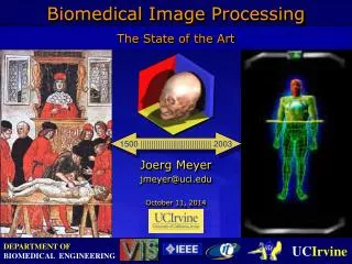
1500 ||||||||||||||||||||||||||||||||| 2003. Biomedical Image Processing. The State of the Art. Joerg Meyer jmeyer@uci.edu December 1, 2014. Biomedical Image Processing. Biomedical Image Processing. (Gundelach Tube, 1898 – 1905) Oak Ridge Associated Universities

E N D
1500 ||||||||||||||||||||||||||||||||| 2003 Biomedical Image Processing The State of the Art Joerg Meyer jmeyer@uci.edu December 1, 2014
(Gundelach Tube, 1898 – 1905) Oak Ridge Associated Universities Health PhysicsInstrumentation Museum Biomedical Image Processing X-Ray (Eberhart’s Manual of High Frequncy Currents, Ch. 10, 1911)
Biomedical Image Processing Week 0 Week 4
Biomedical Image Processing Scaphoid Bone Week 0 Week 4
Biomedical Image Processing Step 1: Incision Marking
Biomedical Image Processing Step 2: Exposure of Fracture Line
Biomedical Image Processing Step 3: Screw Insertion Site
Biomedical Image Processing Before 2 months postop (Images courtesy of: Electronic Textbook of Hand Surgery, http://www.eatonhand.com) Step 4: X-Ray
Biomedical Image Processing Portable System High FrequencyX-Ray Tube (Eberhart’s Manual of High Frequncy Currents, Chap. 10, 1911)
Biomedical Image Processing Scaphoids (1mm) MagneVu 1000(MRI)
Biomedical Image Processing • Problems: • Time • Cost factor • Solution: • Correlate X-ray images with 3-D models • Database (typical bones)
Biomedical Image Processing Scaphoid Bones Various shapes and sizes (Zimmer, 1968)
Biomedical Image Processing Scaphoid Bone Fractures Classification (Herbert)
Biomedical Image Processing A B A - C: Scaphoid view 1 - with forearm pronated 45deg. to view profile of scaphoid & STT joint D - Scaphoid view 2 (ulnar oblique view) showing radioscaphoid joint (from Rockwood & Green) C D
Biomedical Image Processing • Solution: • Superimpose radiographic scan and 3-D model • Select best model from database
Biomedical Image Processing • General Question: • How to combine different modalities?
Visible Human (CT, frozen) Joseph Paul Jernigan
Visible Human (CT, frozen) Slice 1125
Visible Human (MRI) Slice 1125
Visible Human (RGB color) Slice 1125
Image Acquisition Principle of a CT Scanner X-Ray Source X-Ray Source Rotation Object Translation Object Detector Detector a) Translation b) Rotation
Image Acquisition Principle of an MRI Scanner a) Random Spin b) directional magnetic field
Image Acquisition Principle of an MRI Scanner a) Directional Puls (orthogonal) b) Relaxation
Rhesus Monkey Brain • High-resolution large-scale image data RGB image series (real-color, dyed), 5037 x 3871 x 1400, 76 GB (data courtesy of Edward G. Jones, Center for Neuroscience, UC Davis) • Resolution: 2666dpi • Pixel spacing: 9 mm • Enables zoomingdown to the cell level. • Total data size:76 GB