
Human Osteosarcoma Cell Mineralization on Polyarylates and Polycarbonates
Explore cellular response of human osteosarcoma cells to polymer substrates, focusing on mineralization. Study the impact of different polyarylate and polycarbonate substrates on cell behavior. Incorporate variations in polymer chemistry and structure to understand mineralization process. Prepare and analyze polyarylate and polycarbonate-coated glass coverslips. Examine properties affecting flexibility, protein adhesion, and degradation. Evaluate Saos-2 cell mineralization on substrates and draw conclusions. Utilize Alizarin Red-S assay to compare mineralization levels on different substrates.

Human Osteosarcoma Cell Mineralization on Polyarylates and Polycarbonates
E N D
Presentation Transcript
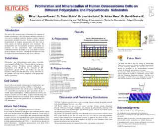
Proliferation and Mineralization of Human Osteosarcoma Cells on Different Polyarylates and Polycarbonate Substrates Milca I. Aponte-Román1, Dr. Robert Dubin2, Dr. Joachim Kohn2, Dr. Adrian Mann1, Dr. David Denhardt3, Departments of 1Materials Science Engineering, and 3Cell Biology & Neuroscience, 2Center for Biomaterials. Rutgers University, The Sate University of New Jersey Introduction Results The goal of this research was to characterize the response of human osteosarcoma cells to polymer substrate variation in vitro and to establish correlation between cellular response and polymer chemistry/structure. In this particularly project mineralization by human osteosarcoma cell line, Saos-2, when grown on various polymers was examined. The biodegradable and biocompatible polymers examined are members of the "polyarylate" and “polycarbonates” combinatorial library developed in the Dr. J. Kohn’s research group at Rutgers University. Alizarin Red-S assay was used to compare the mineralization on each of the substrates. Alizarin, is the main colorant found in the madder plant Rubia tinctorum Polyarylates Poly(DTE suberate) Poly(DTB suberate) Poly(DTH suberate) Poly(DTO suberate) Poly(DTD suberate) Poly(DTE succinate) Poly(DTH succinate) Poly(DTD succinate) Poly(DTE dodecandioate) Poly(DTH dodecandioate) Poly(DTD dodecandioate) Poly(DTH adipate) Poly(DTH sebacate) PLGA poly(DL-lactate co-glycolate) PLA poly(DL-lactate) TCP tissue culture plastic Plate with polycarbonate-coated coverslips after stained with alizarin red-S. Substrates Future Work Polyarylate- and polycarbonate-coated glass coverslips were prepared in Kohn’s lab. Chemico-physical properties affecting polymer flexibility, protein adhesion and degradation were altered by the inclusion of DT, poly(ethylene glycol) (PEG), and Iodine (I2) into the backbone of the poly(DTE carbonate), and by alterations in the pendent chain and diacid componet of the polyarylate family of polymers. This work was done in the Cell Biology & Neuroscience Department as a part of my Integrative Graduate Education and Research Traineeship (IGERT). The main idea was to rotate through the laboratory of my interdisciplinary secondary advisor, Dr. Denhardt, and learn about cell culture. For now on I will be working using a Near Field Scanning Optical Microscope (NSOM) to nanopattern surfaces coated with Self Assembled Monolayers (SAMs). The idea is to functionalize the SAM with bioactive chemicals by using the laser from the NSOM to locally initiate a chemical reaction. B. Polycarbonates Poly(DTE Carbonate) Poly(DTE-co-4% PEG lK Carbonate) Poly(DTE-co-8% PEG lK Carbonate) Poly(DTE-co-10% DT carbonate) Poly(DTE-co-10% DT-co-4% PEG lK Carbonate) Poly(DTE-co-10% DT-co-8% PEG lK Carbonate) Poly(I2 DTE Carbonate) Poly(I2 DTE-co-4% PEG lK Carbonate) Poly(I2 DTE-co-8% PEG lK Carbonate) Poly(I2 DTE-co-10% I2DT lK Carbonate) Poly(I2 DTE-co-10% I2DT-co-4% PEG lK Carbonate) Poly(I2 DTE-co-10% I2DT-c0-8% PEG lK Carbonate) 17. PLGA-resomer 506 (Boehringer Ingelheim) PLLA-resomer L 206 (B. I Chemicals) TCP tissue culture plastic Cell Culture Saos-2 cells were initially cultured on top of polyarylate- and polycarbonate-coated coverslips in HAM’s F-12 medium supplemented with 10% fetal bulvine serum, 10mM Hepes pH 7.5, + penicillin and streptomycin, and + glutamine. At day 7, 10mM -glycerolphosphate, 10nM dexamethasone (DEX), and 50g/mL ascorbic acid were added to induce mineralization, and the addition repeated at every medium change until the end of experiment. Cultures were grown at 37oC in a humidified 5% CO2 atmosphere. Discussion and Preliminary Conclusions • All Saos-2 cultures on polyarylate-coated coverslips formed calcium phospahate mineral with minimal disparity between each other. • All Saos-2 cultures on polycarbonate-coated coveslips formed calcium phosphate mineral. However, low light absorbance was obtained in polycarbonate substrates with %4 and 8% PEG (code #2 and #3 from polycarbonate series). PEG in %8 resulted in poor cell attachment and little spreading. • In contrast, no significant variations were obtained on the polycarbonate cultures where PEG is also present in 4% and 8% (code #10, #11, #14, and #15 from polycarbonate series). When Iodine (I2) is incorporated it seems to suppress the effect of PEG. It is possible that the Iodine may work to suppress the PEG by steric hindrance rather than making the polymer more hydrophobic. • These experiments need to be repeated before firm conclusions can be drawn. Alizarin Red-S Assay Acknowledgments Alizarin red is a dye, which binds selectively to calcium. Every week the medium of one plate was removed and the culture wells were briefly washed with PBS and stained for 10 min with 1% alizarin red-S pH 4.14. Cultures were then rinsed with water and PBS, to remove the stain not associated with calcium mineral deposit. A de-staining procedure was followed using 10% cetylpyridinium chloride in 10nm sodium phospahete pH 7.0 for 15 min in order to measure the optical absorbance. Absorbance measurements were done with a microplate reader with 570nm. • The author would like to express her gratitude to, • Dr. Denhardt from the Department of Cell Biology & Neuroscience, for letting me work in his lab and teaching me about cell culture. • Dr.Robert Dubin, for providing us with the culture plates coated with polycarbonate and polyarylates, and valuable discussion. • Christian Kazanecki,Melissa Weidner and Jennifer Luo for their help in cell culture and for sharing their lab with me. • IGERT for supporting this work.


