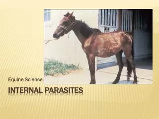
Ruminant Internal Parasites
Ruminant Internal Parasites. By Dr John B Malone, August, 2000. Major Internal Parasites of Ruminants. ABOMASUM INTESTINES Ostertagia(T) Cooperia(T) Strongyloides Haemonchus(T) Trichostrongylus(T) Tapeworm Trichostrongylus(T) Nematodirus(T) Coccidia Bunostomum(S)

Ruminant Internal Parasites
E N D
Presentation Transcript
Ruminant Internal Parasites By Dr John B Malone, August, 2000
Major Internal Parasites of Ruminants ABOMASUM INTESTINES Ostertagia(T) Cooperia(T) Strongyloides Haemonchus(T) Trichostrongylus(T) Tapeworm Trichostrongylus(T) Nematodirus(T) Coccidia Bunostomum(S) Oesophagostomum(S) (T) Trichostrongyloidea (S) Strongyloidea Several trichostrongyles and two strongyles occur in varying proportion as a gastrointestinal nematode “complex”, very often with the additional presence of Strongyloides, tapeworms, and/or coccidia. Internal parasites are enzootic in grazing ruminants
Relative gross size and appearance of Haemonchus (largest), Ostertagia (brownish), Cooperia and Trichostrongylus.
Life Cycle Ostertagia egg Eggs shed by Strongyle-trichostrongyle parasites are similar and are counted by not differentiated by fecal exam, except Bunostomum, which is larger and darker
Life Cycle: Dung Pat Biology Eggs develop to L1, L2 and infective L3 in dung pats at a rate dependent on climatic conditions. This takes about one week under optimum conditions
Development of eggs and free living larvae Eggs passed in feces hatch in 24 hours to rhabditiform L1. In successive molts, rhabitiform L1 and L2, which feed on micro-organisms, develop to filariform infective L3. L3 do not feed (they live on reserves) and are protected from adverse environments by a sheath, the retained cuticle of the L2. Some very resistant species, such as Nematodirus, also retain the L1 cuticle and egg membrane.
L3 picked up at grazing in moisture film on forages in the first 1-2 cm from soil/mulch surface. Most L3 are in moisture film on forage near dung pats and migrate from forage to mulch/soil based on diurnal moisture conditions (ie. dew AM, sun PM)
Rain events disperse larvae to pasture Many mathematical models consider rain events to be a major influence on transmission intensity. ‘Waves’ of larvae become available on wider area of pasture with successive rains
Adult Haemonchus in abomasal lumen at mucus interface Most ruminant nematodes with a direct life cycle have a development time (prepatent period) within the final host of about 3 weeks from infection by L3 to egg shedding adults. L3 exsheath, and molt twice to the L4 and L5 (juvenile adult) stages
Control Programs: A Problem of Population Biology • Rules of Thumb for Parasite Control Based on Epidemiology, Population Dynamics Rule #1 Enzootic Stability < Economic Threshold Rule #2 Assess benefits as a production disease
Enzootic Stability vs Economic Threshhold Diagram illustrating annual control that successfully maintains F. hepatica below economic threshold (40 flukes). Top line represents fluke populations without treatment.
Assess Benefits as a Production Disease Value of parasite control greatest in poorest-managed herds - Reproduction efficiency <90% - Winter loss of body condition of >100lb - Most Rx benefit when “Room to improve” - Leibig’s Law of the minimum = Limits on number and productivity of a population are determined by times of minimum conditions, not times of suitable conditions
Coastal marsh range cattle in good early summer condition. Parasite effects are least at this time.
Common winter condition of coastal marsh cattle. This is the time of maximum adult parasite burdens and greatest effects on host.
“Waiting for a Chinook” C.M. Russell, Musselshoals, Montana
Old Louisiana marsh cow at ‘bottom’ of winter. She is losing her teeth, barely made it through winter. Parasites have major effects in ‘poor’ cattle.
Calf crop weaning weights/quality are influenced by multiple factors, including ‘indirect’ effects of heavy parasitism on < cow mothering ability and milk quantity/quality
Regional Epidemiology Patterns World climate zones affect seasonal dynamics of parasites and other diseases. Epidemiology is unique in each region.
Water Balance in U. S. Warm moist areas favor parasites. Louisiana is in a high risk zone
Modify Control to fit Individual Cattle Operations -Stocking Rate and Pasture Type, Quality Alexandria, LA, Intensive management, Alicia Burmuda/winter rye grass at 1:2 acre stock rate - parasitism exposure is high.
Commercial Cattle Coastal marsh natural range, Cameron, LA at 1:4 to 1:8 acre stocking rate - parasitism exposure is less.
Distribution of beef cow-calf operations. There are three major segments of the cattle industry: Cow-Calf, Stocker, feedlot. Parasite problems vary by industry operation type and location.
Most Southern cow-calf operations sell calf crop as lightweight ‘Stockers’ that graze on small grain pasture (eg winter wheat) until entry to feedlot at about 800 lb. Parasite treatment usually given to stockers at receipt ands at entry to feedlot.
Hypobiosis Adult male Ostertagia and inhibited (hypobiotic) early L4 larvae from the abomasal gastric glands. Hypobiosis allows escape from adverse conditions via climatically adapted genetic strains, although influenced by immunity, crowding effect. Resume develop ‘cue’ unknown.
Evolutionary Competitive Niche Each species has a site both in the host and in the environment where it best survives and competes to the exclusion of other species. In the environment, species have temperature and moisture development adaptations that determine the unique type of habitat and season in which it occurs. In the host, species occupy specific, overlapping regions of the GI tract. The cattle life cycle and parasite life cycle evolved and adapted together. As a rule, there is only ‘one species per niche’.
Parasite Overdispersion: • Egg counts illustrate the ‘overdispersion phenomenon’ in which most worm burdens (and egg shedding) occur in a minority of animals. This is why herd parasite evaluations are done on fecal samples from 10 individuals or a composite of multiple samples, then expressed as eggs per gram (EPG). Herd 1 Herd 2 0 0 2 1 0 7 1 0 0 9 1 2 1 0 3 22 15 5 10 3 %/Mean EPG: 50% / 2.0 90% / 6.2 (16 o 20 in 2 cows) (47 of 62 in 3 cows)
Overdispersion • Majority of parasites in a minority of hosts, with most pasture contamination by a few animals • Related to variable genetic resistance of each individual that evolved to benefit of host and parasite • Advantage = Population Regulation • Insured pasture egg contamination even at low infection rates in periods or places adverse to parasites • With very heavy infection rates, only‘super-susceptibles’ die, not the entire herd; If 10% mortality, >50% of egg shedding stops, down-regulating parasite
Worm Population Turnover and Life Span Cattle in Feedlot Adult parasites are shed after senescence in a few weeks in the absence of re-exposure. (Except inhibited L4.) Ostertagia adults have a life span of 25-56 days, they live longest if low worm burdens and loss is proportional to numbers present. Fluctuation in adult worm burden in host populations is thus related to L3 intake and the proportion that are inhibition prone. Some nematodes are longer lived.
Host - Parasite Equilibrium • Parasitism Host Immunity Nutrition Growing stock in the first and second year of life are most susceptible. Partial immunity builds slowly by long term multiple exposure to a maximum in healthy >4 year-olds, but may be lost during stress or periods of poor nutrition
Forage, cow energy needs and nutritional stress cycle in LA.
Previous slide in relation to nematode and fluke burdens. Maximum nutritional stress, nematode burdens and fluke burdens coincide during the winter period, creating an advantagious time for expanding parasite populations.
Ostertagia(Brown Stomach Worm) Identification Optimum Development Pathology Hypobiosis
Ostertagia cervical papillae are less robust than Haemonchus.
Clinical Ostertagiasis Calf is in poor condition with ‘bottle jaw’ due to hypoproteinemia and anemia. Type I and type II disease have similar effects on animals. Type I is massive direct damage, usually late winter in Louisiana. Type II disease is from sudden emergence of L4 from abomasal wall (Aug-Sept in LA)
“Morrocco leather” due to Ostertagia inhibited larvae. Type II disease is due to massive emergence of inhibited larvae over short period (usually Aug-Oct in LA).
Pathogenesis of Ostertagiasis Type I: Associated with large number of adults, and exposure to large numbers of L3 especially in the first year of life before immunity builds. Development is direct (3-4 weeks). Pathogenic effect is the same as Type II, but no inhibition of L4. Pre-Type II: Up to several thousand inhibited L4 in deep gastric glands (no clinical signs). Type II: Associated with sudden emergence of large numbers of L4, especially yearlings, 2-year olds Anorhexia, green diarrhea, bottle jaw, weight loss. Pathogenesis is by damage to parietal cells (acid production), chief cells (produce pepsinogen), > Inflammatory cells, >bacteria, pepsinogen inactive due to pH 7; Intracellular junctions leak albumen and Na Hypoproteinemia, Abomasal fold edema. Clinical result is anorhexia, green diarrhea, bottle jaw, weight loss
Hypobiosis Plays a Major Role in the Epidemiologic Pattern of Ostertagia
Northern epidemiologic pattern: Adverse climate selects the part of the Ostertagia gene pool that responds to an unknown ‘cue’ to inhibit in winter, emerge in spring. This influences optimum treatment times for control.
Haemonchus (Barber Pole Worm) Identification Optimum Development Pathology Hypobiosis
Haemonchus Note the “barber pole” on live worms grossly visible in abomasum at mucosal surface.
Haemonchus cervical papillae (light and scanning microscopes).
Haemonchus stylet (light and scanning microscopes) assists in blood sucking.
“Bottle Jaw” in merino-x and Targhee sheep due to Haemonchus, the major limiting parasite in Louisiana.