Reactive Oxygen Species
Reactive Oxygen Species. I. Free radicals & ROS Defined II. Sources of ROS III. Oxidative damage in biological systems IV. Antioxidant Defense V. ROS signaling and redox sensitive pathways VI. Oxidative stress and disease VII. Detection methods for ROS & oxidative stress.

Reactive Oxygen Species
E N D
Presentation Transcript
Reactive Oxygen Species I. Free radicals & ROS Defined II. Sources of ROS III. Oxidative damage in biological systems IV. Antioxidant Defense V. ROS signaling and redox sensitive pathways VI. Oxidative stress and disease VII. Detection methods for ROS & oxidative stress
I. Free Radicals & ROS Defined • The Earth was originally anoxic • Metabolism was anaerobic • O2 started appearing ~2.5 x 10⁹ years ago Anaerobic metabolism-glycolysis Glucose + 2ADP + 2PiLactate+2ATP+ 2H2O O2 an electron acceptor in aerobic metabolism Glucose +6O2+ 36ADP + 36Pi 6CO2 +36ATP+ 6H2O
: : . . O:O : : • Ground-state oxygen has 2-unpaired electrons • The unpaired electrons have parallel spins • Oxygen molecule is minimally reactive due to spin restrictions
Basics of Redox Chemistry Term Definition Oxidation Gain in oxygen Loss of hydrogen Loss of electrons Reduction Loss of oxygen Gain of hydrogen Gain of electrons Oxidant Oxidizes another chemical by taking electrons, hydrogen, or by adding oxygen Reductant Reduces another chemical by supplying electrons, hydrogen, or by removing oxygen
Prooxidants R3C.Carbon-centered R3N.Nitrogen-centered R-O.Oxygen-centered R-S.Sulfur-centered • Free Radicals: • Any species capable of independent existence that contains one or more unpaired electrons • A molecule with an unpaired electron in an outer valence shell H2O2Hydrogen peroxide HOCl- Hypochlorous acid O3Ozone 1O2 Singlet oxygen ONOO- Peroxynitrite Men+ Transition metals • Non-Radicals: • Species that have strong oxidizing potential • Species that favor the formation of strong oxidants (e.g., transition metals)
Reactive Oxygen Species (ROS) Radicals: O2.- Superoxide OH. Hydroxyl RO2. Peroxyl RO. Alkoxyl HO2.Hydroperoxyl Non-Radicals: H2O2Hydrogen peroxide HOCl- Hypochlorous acid O3Ozone 1O2 Singlet oxygen ONOO- Peroxynitrite Reactive Nitrogen Species (RNS) Non-Radicals: ONOO-Peroxynitrite ROONO Alkyl peroxynitrites N2O3 Dinitrogen trioxide N2O4 Dinitrogen tetroxide HNO2 Nitrous acid NO2+Nitronium anion NO- Nitroxyl anion NO+ Nitrosyl cation NO2Cl Nitryl chloride Radicals: NO. Nitric Oxide NO2. Nitrogen dioxide
“Longevity” of reactive species Reactive Species Half-life Hydrogen peroxide Organic hydroperoxides ~ minutes Hypohalous acids Peroxyl radicals ~ seconds Nitric oxide Peroxynitrite ~ milliseconds Superoxide anion Singlet oxygen ~ microsecond Alcoxyl radicals Hydroxyl radical ~ nanosecond
Oxidative Stress Antioxidants Prooxidants “An imbalance favoring prooxidants and/or disfavoring antioxidants, potentially leading to damage” -H. Sies
Addition R. + H2C=CH2 R-CH2-CH2. Hydrogen abstraction R. + LH RH + L. Electron abstraction R. + ArNH2 R- + ArNH2.+ Termination R. + Y. R-Y Disproportionation CH3CH2. + CH3CH2. CH3CH3 + CH2=CH2 Radical-mediated reactions
Hydroxyl radical (.OH) O2.-+ Fe3+ O2 + Fe2+ (ferrous) H2O2 + Fe2+ OH- + .OH + Fe3+ (ferric) O2.- + H2O2 OH- + O2 + .OH Fenton Haber-Weiss • Transition metal catalyzed • Other reductants can make Fe2+ (e.g., GSH, ascorbate, hydroquinones) • Fe2+ is an extremely reactive oxidant
Biological Pathways for Oxygen Reduction From: McMurry and Castellion “Fundamentals of general, organic and biological chemistry”
Microsomal Oxidation, Flavoproteins, CYP enzymes Myeloperoxidase (phagocytes) Xanthine Oxidase, NOS isoforms Endoplasmic Reticulum Transition metals Lysosomes Cytoplasm Fe Cu Oxidases, Flavoproteins Peroxisomes Mitochondria Plasma Membrane Lipoxygenases, Prostaglandin synthase NADPH oxidase Electron transport II. Sources of ROS Endogenous sources of ROS and RNS
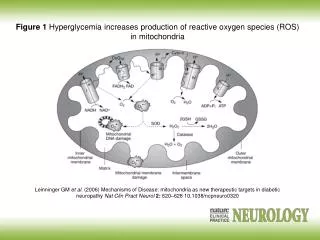
Mitochondria as a source of ROS Mitochondrial electron chain Localization of the main mitochondrial sources of superoxide anion Quinone cycle Turrens, J Physiol, 2003 Chandel & Budinger, Free Radical Biol Med, 2007
Peroxisomes as a source of ROS and RNS Enzymes in mammalian peroxisomes that generate ROS Schader & Fahimi, Histochem Cell Biol, 2004
NADPH oxidase as a source of ROS Present mainly in neutrophils (oxidative burst), but also in many other cell types ANTIOXIDANTS & REDOX SIGNALING Volume 8, Numbers 3 & 4, 2006 Activation of the gp91phox (NOX2) containing NOX complex of phagocytes involves phosphorylation of the cytoplasmic regulator p47phox, with the translocation of the cytoplasmic p47phox, p67phox, and p40phox regulatory components to the plasma membrane to interact with flavocytochrome-b558, which is composed of gp91phox and p22phox. Activation of the complex also involves guanine nucleotide exchange on the GTP-binding protein RAC stimulated by guanine nucleotide exchange factors. Guanine nucleotide exchange on RAC is associated with release of RhoGDI and translocation of RAC from the cytosol to the NOX complex at the plasma membrane.
Prostaglandin H Synthase (PHS) as a source of ROS Co-oxidation of xenobiotics (X) during arachidonic acid metabolism to PGH2 PHS
Cytoplasmic sources of ROS and RNS xanthine oxidase xanthine oxidase Nitric Oxide Synthases (NOS): neuronal nNOS (I) endothelial eNOS (III) inducible iNOS (II) NO•
Lysosome as a source of ROS and RNS Myeloperoxidase undergoes a complex array of redox transformations and produces HOCl, degrades H2O2 to oxygen and water, converts tyrosine and other phenols and anilines to free radicals, and hydroxylates aromatic substrates via a cytochrome P450-like activity
Microsomes as a source of ROS (I) A scheme of the catalytic cycle of cytochrome P450-containing monooxygenases. The binding of the substrate (RH) to ferric P450 (a) results in the formation of the substrate complex (b). The ferric P450 then accepts the first electron from CPR (cytochrome P450 reductase), thereby being reduced to the ferrous intermediate (c). This intermediate then binds an oxygen molecule to form oxycomplex (d), which is further reduced to give peroxycomplex (e). The input of protons to this intermediate can result in the heterolytic cleavage of the O–O bond, producing H2O and the ‘oxenoid’ complex (f), the latter of which then inserts the heme-bound activated oxygen atom into the substrate molecule to produce ROH. In eukaryotic monooxygenases, reactive oxygen species (ROS) are produced by ‘leaky’ branches (red arrows). In one such branch, a superoxide anion radical is released owing to the decay of the one-electron-reduced ternary complex (d). The second ROS-producing branch is the protonation of the peroxycytochrome P450 (e), which forms of H2O2. In addition to these ROS-producing branches, another mechanism of electron leakage appears to be the four-electron reduction of the oxygen molecule with the production of water (Davydov, Trends Biochem Sci, 2001).
Microsomes as a source of ROS (II) Davydov, Trends Biochem Sci, 2001
Exogenous sources of free radicals • Radiation • UV light, x-rays, gamma rays • Chemicals that react to form peroxides • Ozone and singlet oxygen • Chemicals that promote superoxide formation • Quinones, nitroaromatics, bipyrimidiulium herbicides • Chemicals that are metabolized to radicals • e.g., polyhalogenated alkanes, phenols, aminophenols • Chemicals that release iron • ferritin
UVA = 320-400 nm UVB = 290-320 nm UVC = 100-290 nm g-rays UVB 2H2O H2O + e- + H2O* H2O* H + .OH H2O2 OH. + OH. • Primarily a concern in skin and eye • Can also cause DNA damage • Can form singlet oxygen in presence of a sensitizer UV radiation Ionizing radiation • High energy radiation will result in .OH
Quinone redox cycling as a mechanism to generate ROS “Premarin (Wyeth–Ayerst) is the most common drug used for hormone replacement therapy (HRT) and is composed of approximately 50% estrogens and 40% equine estrogens [equilenin (EN) and equilin (EQ)] (9). In vitro experiments have shown that equine estrogens are successively metabolized and are capable of forming various types of DNA damage (9–11) (Figure 1). Like estrogen, EN and EQ are metabolized by cytochrome P450 enzymes (CYP) to their 4-hydroxy and 2-hydroxy forms (9,10). 4-Hydroxyequilenin (4-OHEN) is rapidly auto-oxidized to an o-quinone (4-OHEN-o-quinone) which in turn readily reacts with DNA, resulting in the formation of unique dC, dA and dG adducts (4-OHEN–DNA adducts) with four possible stereoisomers for each base adduct (9,11,12). 4-Hydroxyequilin (4-OHEQ) is also autoxidized to an o-quinone which isomerizes to 4-OHEN-o-quinone. As a result, 4-OHEQ and 4-OHEN produce the same 4-OHEN–DNA adduct (13). Simultaneously, oxidative DNA damage, such as 7,8-dihydro-8-oxodeoxyguanine (8-oxodG), is also generated by reactive oxygen species through redox cycling between the o-quinone of 4-OHEN and its semiquinone radicals (14).” Nucl. Acids Res. (210) 38 (12):e133
Chemicals that form peroxides O3 + Ozone 1O2 + Singlet oxygen
Chemicals that promote O2.- formation NAD(P)H NAD(P)+ Flavoprotein Paraquat radical cation Paraquat O2.- O2
Chemicals that are metabolized to radicals Polyhalogenated alkanes Phenols, aminophenols
+ e- Fenton Chemistry Ferretin Fe2+ • Requires reductant • Promotes .OH formation • Promotes lipid peroxidation in vitro Chemicals that release iron
III. Oxidative Damage in Biological Systems Oxidative stress and cell damage • High doses: • directly damage/kill cells • Low doses/chronic overproduction of oxidants: • activation of cellular pathways • stimulation of cell proliferation • damage to cellular proteins, DNA and lipids
Classic lipid peroxidation • Initiation • LH + X• L• + XH • 2. Propagation • L• + O2 LOO• • LOO• + LH L• + LOOH • 3. Termination • 2 LOO• non-radical products • L• + LOO• non-radical products • L• + L• non-radical products Catalyzed by metals LOOH + Fe2+ OH- + LO. + Fe3+
Consequences of lipid peroxidation • Structural changes in membranes • alter fluidity and channels • alter membrane-bound signaling proteins • increases ion permeability • Lipid peroxidation products form adducts/crosslinks with non lipids • e.g., proteins and DNA • Cause direct toxicity of lipid peroxidation products • e.g., 4-hydroxynonenal toxicity • Disruptions in membrane-dependent signaling • DNA damage and mutagenesis
Protein targets for ROS Cysteine Methionine Tyrosine Oxidized proteins and amino acids found in biological systems Histidine Tryptophan
Consequences of protein thiol oxidation • Oxidation of catalytic sites on proteins • loss of function/abnormal function • BUT(!): sometimes it is gain in function! • Formation of mixed sulfide bonds • Protein-protein linkages (RS-SR) • Protein-GSH linkages (RS-SG) • Alteration in 2o and 3o structure • Increased susceptibility to proteolysis
DNA oxidation products 8-hydroxyguanine 8-hydroxyadenine 2-hydroxyadenine 5,8-dihydroxycytosine thymidine glycol 5-hydroxymethyluracil
Oxidation of deoxyribose (DNA backbone) Strand Breaks O2 + B + Apurinic/apyriminic sites Aldehyde products
Consequences of DNA oxidation • DNA adducts/AP sites/Strand breaks • mutations • initiation of cancer • Stimulation of DNA repair • can deplete energy reserves (PARP) • imbalanced induction of DNA repair enzymes • induction of error prone polymerases • activation of other signaling pathways
IV. Antioxidant Defense Defenses against Prooxidants • Prevention of prooxidant formation • Interception of prooxidants • Breaking the chain of radical reactions • Repair of damage caused by prooxidants ANTIOXIDANT: a substance that is able, at relatively low concentrations, to compete with other oxidizable substrates and, thus, to significantly delay or inhibit the oxidation of other substrates
Prevention of prooxidant formation Physical prevention: Behavioral: - avoidance Barriers: - organismal level - organ level - cellular level Biochemical prevention: Control of prooxidant molecules: - transition metal chelators - catalytic control of O2 reduction Control of prooxidant enzymes: - blockade of stimuli - inhibition of enzymes
Examples of preventative ‘antioxidants’ Anti-inflammatory agents Nitric oxide synthase inhibitors Metal chelators: - Metallothionein - Transferrin - Lactoferrin NADPH oxidase inhibitors Xanthine oxidase inhibitors
Interception of prooxidants ‘Classical’ antioxidant: Intercepts species, once formed Excludes from further damaging activity Transfers species from critical parts of cell Important considerations for interception reactions: Speed of reaction (rate constant) Concentration of intercepting species in vivo Is reaction truly a detoxication pathway? Is reaction catalytically recyclable?
Chain breaking antioxidants Example of radical chain-reaction: lipid peroxidation ROO• (peroxyl radicals) are often the chain-carrying radicals Chain-breaking oxidants act by reacting with peroxyl radicals: “Donor” antioxidants (tocopherol, ascorbate, uric acid,…) LOO• + TOH LOOH + TO• “Sacrificial” antioxidants (Nitric oxide): LOO• + NO• LOONO • Good chain-breaking antioxidant: • both ANT and ANT• should be relatively UNreactive • ANT• decays to harmless products • does not add O2 to make a new peroxyl radical • is renewed (recycled)
a) Initiation of the peroxidation by an oxidizing radical, X', by abstraction of a bis-allylic hydrogen, forming a pentadienyl radical. b) Oxygenation to form a peroxyl radical and a conjugated diene. c) The perolcyl radical moiety partitions to the water-membrane interface where it is poised for repair by tocopherol. d) The tocopheroxyl radical can be repaired by ascorbate. e) Tocopherol has been recycled by ascorbate; the resulting ascorbate radical can be recycled by enzyme systems. The enzymes phospholipase AZ (PLAZ), phospholipid hydroperoxide glutathione peroxidase (PhGPx), glutathione peroxldase (GPx), and fatty acyl-coenzyme A (FA-CoA), cooperate to detoxify and repair the oxidized fatty acid chain of the phospholipid.
Cellular antioxidants Small Molecules -Water soluble: glutathione, uric acid, ascorbate (Vit. C) -Lipid soluble: a-tocopherol (Vit. E), b-carotene, coenzyme Q Proteins -Intracellular: SOD (I and II), glutathione peroxidase, catalase -Cell membrane: SOD (III), ecGPx, plasma proteins (e.g. albumin) -Extracellular: phospholipid hydroperoxide GPx (PHGPx) See additional information on antioxidant enzymes in handout material
‘Antioxidant Network’ Catalytic maintenance of antioxidant defense Non-scavenging enzymes (re-reduce antioxidants) Dependence on energy status of cell Glucose most important ‘antioxidant’ Catalytic reduction of peroxides Catalytic reduction of lipid radicals
Repair of damage caused by prooxidants Protection not perfect Repair of damaged products proteins and lipids -rereduction and degradation DNA -repair enzymes Cell death (apoptosis/necrosis)
V. ROS signaling and redox sensitive pathways Environmental factors Endogenous mediators Normal metabolism
Agonist Receptor Primary effectors (enzymes or channels) Second messenger(s) Secondary effectors (enzymes, other molecules) Signal transduction: Regulated sequence of biochemical steps through which a stimulant conveys a message, resulting in a physiologic response Oxidants can act to modify signal transduction
How free radicals can be involved in signaling? • Heme oxidation • Oxidation of iron-sulfur centers in proteins • Changes in thiol/disulfide redox state of the cell • Change in conformation change in activity • Oxidative modification of proteins: degradation, loss of function, or gain of function • Oxidative modification of DNA: activation of repair, and/or apoptosis • Oxidative modification of lipids: disruption of membrane-associated signaling, DNA damage, and formation of protein adducts