Download
1 / 41
410 likes | 580 Vues
Understanding Disseminated Intravascular Coagulation (DIC) “An Oncologic Emergency”. Deborah Kwasneske RN, BSN, OCN MSN 621, Spring 2006 Alverno College Email: debbiern@wi.rr.com. Welcome.

E N D
Understanding Disseminated Intravascular Coagulation (DIC) “An Oncologic Emergency” Deborah Kwasneske RN, BSN, OCN MSN 621, Spring 2006 Alverno College Email: debbiern@wi.rr.com
Welcome This tutorial is self guided by using buttons to move about screens. You can return to the main screen at anytime by using the home button. The arrow buttons allow you to move forward or back. To return to previous spot you may use the return button To navigate through different topics of this tutorial, use the button. There are multiple hyperlinks to outside learning sources to help with the understanding of DIC . To get back to DIC tutorial use back button on links. For the case study answers, just click on the question for answers to appear. You can return to the case study using the return key.
Understanding Disseminated Intravascular Coagulation (DIC) “An Oncologic Emergency” Introduction DIC What isDIC Genetics Inflammation Pathophysiology Treatments Case Study Glossary References
Objectives • Understand the pathophysiology of Disseminated Intravascular Coagulation (DIC) • Identify risk factors and etiology of DIC • Describe the signs and symptoms of DIC • Identify treatment modalities for DIC • Define, identify and understand Acute vs Chronic DIC • Develop understanding of diagnosing DIC (lab interpretations)
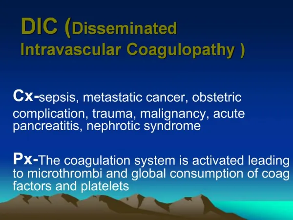
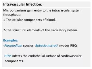
What is DIC? • Is considered an “acquired bleeding disorder” • Is not a disease entity but an event that can accompany various disease processes • Is an alteration in the blood clotting mechanism:abnormal acceleration of the coagulation cascade, resulting in thrombosis • As a result of the depletion of clotting factors, hemorrhage occurs simultaneously • Is a Paradoxical Clinical Presentation“clotting and hemorrhage” (Porth, C.M. (2004) Essentials of Pathophysiology) & (Otto, S. (2001). Oncology Nursing)
Pathophysiology In DIC, a systemic activation of the coagulation system simultaneously leads to thrombus formation (compromising blood supply to various organs) and exhaustion of platelets and coagulation factors (results in hemorrhage). This is a disruption of body homeostasis. (Porth, 2001)
Thrombosis-brief period of hypercoagulability Coagulation cascade is initiated, causing widespread fibrin formation Microthrombi are deposited throughout he microcirculatory Fibrin deposits result in tissue ischemia, hypoxia, necrosis Leads to multi organ dysfunction (Porth, 2004) & (Otto, 2001) Fibrinolysis-period of hypocoagulability (the hemorrhagic phase) Activates the complement system Byproducts of fibrinolysis (fibrin/fibrin degradation products(FDP)) further enhance bleeding by interfering with platelet aggregation, fibrin polymerization, & thrombin activity Leads to Hemorrhage Pathophysiology
Pathophysiology (Porth, 2004)
Risk Factors and Etiology • Almost always a secondary event from activation of one of the coagulation pathways • Underlying pathology creates a triggering event: Either- • endothelial tissue injury (Extrinsic) • blood vessel injury (Intrinsic) (Porth, 2004)
Extrinsic (endothelial) Shock or trauma Infections ( gram positive and gram negative sepsis, aspergillosis) Obstetric complications (eclampsia, placenta abruptio, fetal death syndrome) Malignancies: APML, AML, cancers of the lung, colon, breast, prostate) (Porth, 2004) & (Otto, 2001) Intrinsic (blood vessel) Infectious vasculitis (certain viral infections, rocky mountain spotted fever) Vascular disorders Intravascular hemolysis (hemolytic transfusion reactions) Miscellaneous: snakebite, pancreatitis, liver disease Pathologic Pathways
Oncology Related DIC • Usually related to: • Disease process -APML( acute promyelocytic leukemia) -mucin-secreting adenocarcinomas • Treatment of cancer -chemotherapy -radiation • Concomitantly with sepsis -10-20% with gram-negative (Otto, S. (2001). Oncology Nursing)
Clinical Features • Onset maybe Acute or Chronic • Acute DIC • Develops rapidly over a period of hours • Presents with sudden bleeding from multiple sites • Treated as a medical emergency • Chronic DIC • Develops over a period of months • Maybe subclinical • Eventually evolves into an acute DIC pattern (Otto, 2001)
Signs and Symptoms Most common sign of DIC is bleeding -manifested by ecchymosis, petechiae,and purpura -bleeding from multiple sites either oozing or frank bleeding -cool and or mottled extremities may be noted -dyspnea and chest pain if pleura and pericardium involvement -hematuria (Porth, 2004) & (Otto, 2001)
Genetic Component None associated with DIC BUT------- Genetic mutations to the clotting cascade can lead to coagulation disorders: Hemophilla A and B von Willebrand Factor Impaired Synthesis of Coagulation Factors tutorial on homeostasis http://alverno.edu.bownep
Inflammation and DIC • Sepsis is usually underlying process for development • Endotoxins associated with sepsis activate factors and initiate coagulation. • Bacteria, virus also trigger the clotting cascade. • Sepsis activates complement cascade. http://tutorial on inflammation P.Bowne, Alverno College
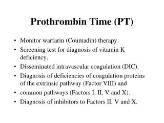
Test Platelet count Fibrin degradation product (FDP) Factor assay Prothrombin time (PT) Activated PTT Throbimn time Fibrinogen D-dimer Antithrombin Abnormality Decreased Increased Decreased Prolonged Prolonged Prolonged Decreased Increased Decreased(Otto,2001) Diagnosis/Lab Findings
Treatment Modalities • Treat the underlying cause • Provide supportive management of complications • Support organ function • Stop abnormal coagulation and control bleeding by replacement of depleted blood and clotting components (FFP,Platelets,PRBC) • Medications can be used and choice depends on the patient’s condition (Heparin, Antithrombin III (ATIII), Fibrinolytic inhibitors) (Porth, C.M. (2004) Essentials of Pathophysiology) & (Otto, S. (2001). Oncology Nursing)
Case Study/Post Test D.G.,a 48-year-old, is 30 days post matched unrelated allogenic stem cell transplant for Acute Myelegenous Leukemia. His Lab work is as follows: WBC 480 Sodium 130 ANC 120 Potassium 3.7 Plts 35 BUN 16 Hct 27 Creatinine 1.4 Hgb 10 (a) Based on the above lab values, what risk factors does D.G. have? The next morning after labs are drawn, D.G. developed a fever of 101.2 and BP fell to 98/60 (normally 130/76). (b)What may be the cause of the fever and low BP? (c)What interventions should you as primary RN take at this time?
Case Study cont. D.G. is started on Vanco and Primaxin and given 1 liter fluid bolus over 2 hours. Blood cultures were drawn prior to antibiotic RX. His fever decreased to 99.8 and BP increased to 108/70. Overnight D.G. again became febrile to 102.4 with drop in BP to 70’s/50’s. Labs were drawn and fluids started at 500cc/hr. Labs values were significant: ANC 100 Plts 20 Hct 9 BUN 35 Hgb 24 Creat 2.8 (a)What do the above lab values say about D.G. status? (b)What interventions should be taken?
Case Study cont. MD orders 2 units of PRBC to be infused over 4 hours. During infusion BP increased to 90/48 and he continues to be febrile at 101.0. D.G. is becoming more confused, complaining of GI pain and cramping, developing a moist cough with rapid respiratory rate. (a)What maybe causing the new symptoms? (b)What interventions do you take at this time? (c)What maybe the underlying process? What else is he at risk for?
Case Study cont. O2 sats on RA measure 84% and lung sounds are coarse throughout. STAT ABG’s and CXR are done. X-ray results show diffuse consolidation and the ABGs demonstrate respiratory acidosis with hypoxemia. D.G. is placed on oxygen, O2 sats improve to 91%. Urine and stool output at this time test heme positive. You also note he has small lower extremity bruising and scattered petechia. A bronchoscophy is ordered and performed. Results show diffuse alveolar hemorrhage. (a) Based on D.G. history and the current clinical picture, what are potential causes for symptoms (GI, Respiratory, GU)? (b) What information would be useful at this time for diagnosis?
Case Study Cont. MD orders CBC and coagulation screen. The results are as follows: WBC <100 PT 34 sec ANC 0 PTT 72 Plts 12 Fibrinogen <100 Hct 23 FSP >1000 Hgb 9 D-dimer >500 (c)What secondary process is possibly occurring based on above values? (d)What interventions are appropriate? What should be the focus of your assessments?
Case Study Cont. D.G. Continues to be hypotensive. Lung sounds remain course, hemoptysis develops and CXR continues with diffuse consolidation. Urine and stool remain heme positive. After transfusion, labs redrawn. Values as follows: Plts 22 Pt 28 Hct 26 PTT 54 Hgb 10 Fibrin <100 FSP >1000 (a)What is the results of treatment based on above values? (b)What interventions should be done?
Case Study cont. D.G. status continues to deteriorate and needed to be intubated 2 days after developing DIC. The sepsis remained unresolved and he went into acute renal failure. Family decided not to dialyze due to the multi-organ involvement. D.G. expired 2 days later from the gram neg sepsis and secondary DIC.
Answers for Case Study (a)Risk for infection, renal insufficiency, neutropenia, bleeding based on lab values. WBC low along with the ANC makes patient more susceptible to infection. The BUN and creatinine are elevated leading to possible renal problems
Answers for Case Study • Possible septic shock as a result of an underlying infection. Neutropenic patients are at high risk for development of infections. (c) Patient will need blood cultures and labs drawn, a fluid challenge is needed to help with low BP’s. Will need to be started on antibiotics. Patient will need to be monitored closely, VS, I&O’s …
Answers for Case Study • D.G.’s labs cont to show anemia, neutropenia, renal insuffiency/failure. BUN and creatinine have increased and the H&H has falling since previous draw. The ANC conts to drop. • Fluids, PRBC transfusion, monitoring of urine output and VS are needed at this time. The transfusion and fluids will help with hypotension and anemia.
Answers for Case Study • Pt possibly developing hypoxemia as a result of the infection process, hypotension. • Need to monitor O2 sats and apply oxygen. Need to get orders for CXR and cont to monitor Vital signs closely.
Answers for Case Study • Patient maybe developing pneumonia and is at an increased risk for ARDS (acute respiratory distress syndrome). The moist cough with rapid resp. rate is good indication of pulmonary involvement. The patient also has increased risk of developing DIC based on the clinical presentation and risk factors presented.
Answers for Case Study (a) The clinical presentation leads to the diagnosis of DIC (effects every system of the body); Lungs- hypoxemia, hemorrhage, tachypnea Cardiac- acidosis, tachycardia GI- cramping, abdominal pain Renal- hematuria Integument- bruising, petechiae Mental status changes- confusion
Answer for Case Study • A DIC panel results would be helpful at this time. Lab abnormalities along with clinical presentation is used to confirm a diagnosis of DIC. If abnormal results are obtained for PT, PTT, platelets, and fibrinogen, then the D-dimer and FDPs levels are used to confirm DIC...FDPs abnormal in 75% and D-dimer in 95%. (Otto, 2001)
Answers for Case Study • Based on the lab values, DIC is confirmed for the patient. The Platelet count is decreased, the fibrinogen level is decreased, PT and PTT levels increased and prolonged, FDPs level is increased and the D-dimer is increased (usually together, levels are 100% specificity and sensitive). If a factor assay was done one would expect the levels to show a decrease in factors VI, VIII, and IX (Otto, 2001)
Answers for Case Study • The immediate goal for the overall management of DIC is the treat the underlying disorder and to stop the patient from actively bleeding and clotting with the need to give transfusions and anticoagulation therapy if needed.* Focus of assessments is to monitor for more bleeding and changes. *Heparin therapy has met with controversy for the treatment of DIC (Porth, 2004) & (Otto, 2001)
Answers for Case Study • Based on the lab values, DG shows slight improvement. The PT and PTT numbers are better, platelet level has increased. • The patient will cont to need to be monitored very closely. Will need to cont with treatment, monitor lab values frequently. Patient remains critically ill.
References Otto, Shirley E. (2001). Oncology Nursing. Mosby: St. Louis. Porth, Carol M. (2004). Essentials of Pathophysiology: Concepts of Altered Health States. Lippncott Williams & Wilkins: Philadelphia. Web Sites: Pat Bowne, faculty Alverno College Milwaukee Wis.
Glossary Complement-a heat-labile cascade system of at least 20 glycoproteins in normal serum that interacts to provide manu of the effector functions of humoral immunity and inflammation, including vasodilation and increases of vascular permeability, facilitation of phagocyte activity and lysis of certain foreign cells. F’s (factors) of Coagulation:factors essential to normal blood-clotting, whose absence, diminution, or excess may lead to abnormality of clotting mechanisms. There are 12 Factors-designated by Roman Numerals. (Porth, 2004) & (Otto, 2001)
Glossary Fibrin- an insoluble protein that is essential to clotting of blood, formed from fibrinogen by action of thrombin Fibrinogen- a coagulation factor I, a glycoprotein; administered to increase the coagulability of the blood Fibrinolysin- plasmin, a preparation of proteolytic enzyme formed from profibrinolysin(plasminogen); to promote dissolution of thrombi Hemostasis-the condition in which the external and internal environment of a cell remains relatively constant Thrombin- an enzyme resulting from activation of prothrombin, which catalyzes the conversion of fibrinogen to fibrin. Thrombosis-formation,development or presences of a thrombus Thrombus-an aggregation of blood factors, primarily platlets and fibrin with entrapment of cellular elements, frequently causing vascular obstruction at the point of formation. (Porth, 2004) & (Otto, 2001)
Glossary Activated PTT- aPTT tests the intrinsic and common pathways D-dimer- an antigen formed as a result of plasmin lysis of cross-linked fibrin clots, documents the presence of thrombin Fibrin degradation product(FDP)- degradation products increase as plasmin biodegrades fibrinogen and fibrin,levels are elevated in 85-100% of patients with DIC Prothrombin Time(PT)- tests the extrinsic and common pathways (Porth, 2004) & (Otto, 2001)
Glossary Blood Components: used to correct abnormal homeostasis. Used to correct the clotting deficiencies caused by the consumption of blood components during the DIC process. -Platelets: contain platelet factor III, which functions as a mechanical plug -Fresh frozen plasma (FFPs): used for volume expansion and contains clotting factors V,VIII,XIII and antithrombinIII. (Otto, S. (2001) Oncology Nursing.)
Glossary Blood Components cont’d: Packed Red Blood Cells (PRBC’s): used to increase RBC’s and clotting factors. Used instead of whole blood to help with fluid overload and reduce development of antibodies. Cryoprecpitate: contains fibrinogen and factor VIII. (Otto, S. (2001). Oncology Nursing)
Thank you This completes the DIC tutorial. Any and all comments regarding your learning experience are greatly appreciated. Please send any correspondence to my email address listed below: debbiern@wi.rr.com