Download
1 / 17
210 likes | 586 Vues
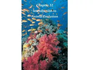
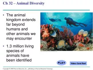
Chapter 32 An Introduction to Animal Diversity. What is an animal? Multicellular, heterotrophic eukaryote – ingestion Structural support from structural proteins – NOT cell walls Nervous tissue & muscle tissue for impulse conduction & movement

E N D
Chapter 32 An Introduction to Animal Diversity • What is an animal? • Multicellular, heterotrophic eukaryote – ingestion • Structural support from structural proteins – NOT cell walls • Nervous tissue & muscle tissue for impulse conduction & movement • Sexual reproduction with motile sperm swimming to non-motile egg • How did animals evolve? • - Current animal development
Cleavage Zygote Eight-cell stage Figure 32.2 Early embryonic development in animals (layer 1) Cleavage – cell division w/out cytokinesis - More cells but same total volume – no cell growth
Blastocoel Cleavage Cleavage Cross section of blastula Zygote Eight-cell stage Blastula Figure 32.2 Early embryonic development in animals (layer 2) Blastula – hollow ball of cells -coel – opening or cavity
Blastocoel Cleavage Cleavage Cross section of blastula Zygote Eight-cell stage Blastula Blastocoel Endoderm Ectoderm Gastrula Gastrulation Blastopore Figure 32.2 Early embryonic development in animals (layer 3) Gastrulation – movement of cells to form 2 layers Blastopore – opening where cells move into Ectoderm – outside layer Endoderm – inside layer
Chapter 32 An Introduction to Animal Diversity • What is an animal? • Multicellular, heterotrophic eukaryote – ingestion • Structural support from structural proteins – NOT cell walls • Nervous tissue & muscle tissue for impulse conduction & movement • Sexual reproduction with motile sperm swimming to non-motile egg • How did animals evolve? • Current animal development • Current hypothesis
Digestive cavity Somatic cells Reproductive cells Hollow sphereof unspecialized cells (shown in cross section) Colonial protist, an aggregate of identical cells Beginning of cell specialization Infolding Gastrula-like “protoanimal” Figure 32.4 One hypothesis for the origin of animals from a flagellated protist
Radial Bilateral Chapter 32 An Introduction to Animal Diversity • What is an animal? • How did animals evolve? • How are animals grouped & characterized? • Body plans • Symmetry • None (sponge) • Radial • multiple central axis “cuts” provide mirror images • Only have top & bottom • Bilateral • Only 1 central axis “cut” provides a mirror image • Has top, bottom, left & right
H-W Lab • All 6 sections • Personal Acct • Data for all 3 Cases including NEW p & q • Discussion questions for all 3 Cases • 6 practice problems at the end • Not Exercise 8A or Case IV • Protist ID Lab • -All 6 sections • -Pre-lab from in-class notes • -Personal acct • -include characteristics that you keyed out • -name of organism • -drawing of organism
Chapter 32 An Introduction to Animal Diversity • What is an animal? • How did animals evolve? • How are animals grouped & characterized? • Body plans • Symmetry & TISSUES • None (sponge) • Radial • multiple central axis “cuts” provide mirror images • Only have top & bottom • Diploblastic – 2 cell layers • Ectoderm • Endoderm • Bilateral • Only 1 central axis cut provides a mirror image • Has top, bottom, left & right • Triploblastic – 3 cell layers • Ectoderm & endoderm • Mesoderm – in between ecto- & endoderm • Cephalization – movement of sensory equipment towards the • anterior end of the organism – associated with movement
Chapter 32 An Introduction to Animal Diversity • What is an animal? • How did animals evolve? • How are animals grouped & characterized? • Body plans • Symmetry & tissues • Body cavities • Acoelomates – without a body cavity • Pseudocoelomates – “false body cavity” – cavity not completely • lined with tissue derived from mesoderm • Coelomates – body cavity completely lined with tissue • from mesoderm
Coelom Body covering (from ectoderm) (a) Coelomate. Coelomates such as annelids have a true coelom, a body cavity completely lined by tissue derived from mesoderm. Tissue layer lining coelom and suspending internal organs (from mesoderm) Digestive tract (from endoderm) Body covering (from ectoderm) (b) Pseudocoelomate. Pseudocoelomates such as nematodes have a body cavity only partially lined by tissue derived from mesoderm. Muscle layer (from mesoderm) Pseudocoelom Digestive tract (from ectoderm) Body covering (from ectoderm) Tissue- filled region (from mesoderm) (c) Acoelomate. Acoelomates such as flatworms lack a body cavity between the digestive tract and outer body wall. Digestive tract (from endoderm) Figure 32.8 Body plans of triploblastic animals
Chapter 32 An Introduction to Animal Diversity • What is an animal? • How did animals evolve? • How are animals grouped & characterized? • Body plans • Symmetry & tissues • Body cavities • Developmental plan • Protostome • Deuterostome
Protostome development (examples: molluscs, annelids, arthropods) Deuterostome development (examples: echinoderms, chordates) (a) Eight-cell stage Eight-cell stage Cleavage. In general, protostome development begins with spiral, determinate cleavage. Deuterostome development is characterized by radial, indeterminate cleavage. Spiral and determinate Radial and indeterminate (b) Coelom Coelom formation. Coelom formation begins in the gastrula stage. In protostome development, the coelom forms from splits in the mesoderm (schizocoelous development). In deuterostome development, the coelom forms from mesodermal outpocketings of the archenteron (enterocoelous development). Archenteron Coelom Mesoderm Blastopore Mesoderm Blastopore Schizocoelous: solid masses of mesoderm split and form coelom Enterocoelous: folds of archenteron form coelom (c) Mouth Anus Fate of the blastopore. In protostome development, the mouth forms from the blastopore. In deuterostome development, the mouth forms from a secondary opening. Digestive tube Mouth Anus Anus develops from blastopore Mouth develops from blastopore Figure 32.9 A comparison of protostome and deuterostome development
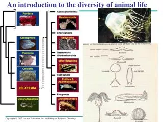
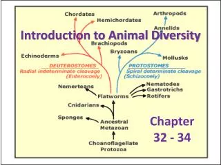
Rotifera Porifera Cnidaria Annelida Mollusca Chordata Nemertea Nematoda Phoronida Ectoprocta Arthropoda Ctenophora Brachiopoda Echinodermata Platyhelminthes “Radiata” Deuterostomia Protostomia Bilateria Eumetazoa Metazoa Ancestral colonial flagellate Figure 32.10 One hypothesis of animal phylogeny based mainly on morphological and developmental comparisons
Rotifera Cnidaria Silicarea Chordata Annelida Calcarea Mollusca Nemertea Nematoda Phoronida Ectoprocta Arthropoda Ctenophora Brachiopoda Echinodermata Platyhelminthes “Radiata” Deuterostomia Lophotrochozoa Ecdysozoa “Porifera” Bilateria Eumetazoa Metazoa Ancestral colonial flagellate Figure 32.11 One hypothesis of animal phylogeny based mainly on molecular data
Figure 32.12 Ecdysis Ecdysozoa – secrete an exoskeleton and molt Nematoda & Arthropoda
Apical tuft of cilia Mouth 100 m Anus (b) Structure of trochophore larva (a) An ectoproct, a lophophorate Figure 32.13 Characteristics of lophotrochozoans