Download
1 / 96
1.06k likes | 1.44k Vues
The Digestive System and Body Metabolism. Chapter 14. Anatomy of the Digestive System. The digestive system is broken down into two main groups: Alimentary canal Responsible for the actual digestion and absorption of foods Accessory digestive organs Assist in the digestive breakdown process.

E N D
The Digestive Systemand Body Metabolism Chapter 14
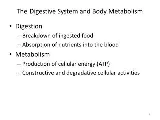
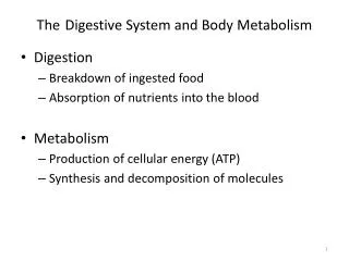
Anatomy of the Digestive System • The digestive system is broken down into two main groups: • Alimentary canal • Responsible for the actual digestion and absorption of foods • Accessory digestive organs • Assist in the digestive breakdown process
Organs of the Alimentary Canal • Also called the Gastrointestinal Tract (GI) • A continuous coiled, hollow muscular tube that winds through the ventral body cavity and is open at both ends. • Organs consist of the: • Mouth • Pharynx • Esophagus • Stomach • Small intestine • Large intestine • Anus • In a cadaver the GI tract is approximately 9 m (30 ft) long but in a healthy living adult is considerably shorter because of its constant muscle tone. • Food in this area is considered “outside” the body because it only has contact with cells that line the GI tract and is open to the external environment on both ends.
Organs of the Alimentary CanalGI Tract • Mouth/Oral cavity: • Mucus lined cavity where food enters the digestive tract. • Lips (Labia): protect the anterior opening • Cheeks: form the lateral walls • Hart palate: forms the anterior roof of the mouth • Soft Palate: forms the posterior roof of the mouth
Organs of the Alimentary CanalGI Tract • Mouth con’t: • Uvula: fleshy fingerlike projection of the soft palate that extends downward. • Vestibule: the space between the lips and cheeks externally and between the teeth and gums internally • Oral cavity proper: area where teeth are contained. • Tongue: muscle occupying the floor of the mouth. Attaches to the hyoid bone and the styloid process of the skull • Frenulum: fold of mucous membrane which secures the tongue to the floor of the mouth and limits posterior movement.
Disorders within the Mouth • “Tongue Tied”: Children born with an extremely short frenulum have distorted speech due to restricted tongue movement. This can be corrected surgically • Tonsillitis: inflammation of the tonsils. • Tonsils are found at the posterior end of the oral cavity and are a mass of lymphatic tissue • Two pairs of tonsils within the mouth: Palatine tonsils, Lingual tonsils • Are a part of the body’s defense mechanism and when they become inflamed will block the pharynx making swallowing painful and difficult.
Organs of the Alimentary CanalGI Tract • Pharynx: • Oropharynx: • Posterior to the oral cavity • Laronygopharynx: • Continuation of the oropharynx which turns into the esophagus which lies below it • Both are passageways for food, air and fluids
Organs of the Alimentary CanalGI Tract • Esophagus: • Runs from the pharynx through the diaphragm to the stomach and is approx. 10 inches long • Passageway for food to reach the stomach
Walls of the Alimentary Canal Organs • The alimentary organ walls consists of four basic tissue layers/tunics: • Mucosa • Submucosa • Muscularis Externa • Serosa
Walls of the Alimentary Canal Organs • Mucosa: • Innermost layer • Moist membrane lining the cavity of the organ • Consists mostly of surface epithelium and a small amount of connective and smooth muscle tissue
Walls of the Alimentary Canal Organs • Submucosa: • Found just beneath the mucosa • Made up of a soft connective tissue layer which contains blood vessels, nerve endings, lymph nodes and lymphatic vessels
Walls of the Alimentary Canal Organs • Muscularis Externa: • Muscle layer made up of an inner circular layer and an outer longitudinal layer of smooth muscle cells.
Walls of the Alimentary Canal Organs • Serosa: • Outermost layer of the wall • Consists of a single layer of flat serous fluid producing cells creating the • Visceral peritoneum which continues into the • Partial peritoneum which lines the abdominopelvic cavity by a membrane extension called the mesentery.
Nerve Plexuses of theAlimentary Canal • The Alimentary canal contains two important nerve plexuses: • Submucosal nerve plexus • Myenteric nerve plexus • These are networks of nerve fibers which are part of the autonomic nervous system and help regulate the mobility and secretory activity of the GI tract organs.
The Stomach • The stomach is a c-shaped organ located on the left side of the abdominal cavity almost hidden by the liver and diaphragm • Cardioesophageal Sphincter: located at the part of the stomach closest to the heart. Area where food enters the stomach from the esophagus. • Body: Middle portion of the stomach • Pylorus: funnel shaped portion of the stomach located on the inferior end of the stomach and continues as part of the wall of the small intestine by the pyloric sphincter
The Stomach • The stomach is approximately 10 inches long but its diameter can change depending on the amount of food within it. • If the stomach is full it can hold approximately 1 gallon of food. When it is empty it collapses in on itself and becomes a mass of folds called rugae.
The Stomach • The lateral surface of the stomach is more convex and referred to as the greater curvature. • The Medial surface is more concave and referred to as the lesser curvature. • Lesser Omentum: a double layer of peritoneum which extends from the lesser curvature to the liver • Greater Omentum: another extension of the peritoneum which drapes downward and covers the abdominal organs and attaches to the posterior body wall of the stomach • The greater omentum is covered with fat which helps to insulate, cushion and protect the organs and also contains lymph nodes and defensive cells of the immune system.
The Stomach • The stomach is a storage area for food as well as a site for food breakdown. • The walls of the muscularis externa help to move food along the digestive tract and also to mix and break down the food into smaller fragments. • The chemical breakdown of foods also occurs in the stomach by producing excessive amounts of mucus and secreting gastric juice from the gastric glands which are found in hollow pits throughout the stomach • Chief cells produce a protein-digesting enzyme made up of pepsinogens • Parietal cells produce hydrochloric acid which makes stomach contents acidic and activates the enzymes. • Mucus neck cells produce a sticky alkaline mucus that clings to the stomach walls protecting it from the acids and digestive enzymes • Chyme: a heavy cream substance that is created from the processing of foods in the stomach. It also allows it to move through the small intestine.
Complications of the Stomach • Peritonitis: • Infection of the peritoneum causing the membranes to stick together around the infection site helping to seal off and localize the infection.
The Small Intestine • The body’s major digestive organ • Passageway for foods that are usable to be moved into the cells of the body • The small intestine is a muscular tube extending from the pyloric sphincter, on the bottom of the stomach, to the ileocecal valve (the beginning of the large intestine) • It is the longest section of the alimentary canal and averages 6 ft in length
The Small Intestine • The small intestine has three subdivisions: • Duodenum: curves around the head of the pancreas and is approximately 10 inches long • Jejunum: about 8 feet long and extends from the duodenum to the ilium • Ilium: about 12 feet long. The terminal portion of the small intestine and joins the large intestine at the ileocecal valve.
The Small Intestine • The small intestine can only process small amounts of food at one time and therefore depends on the pyloric sphincter to control the movement of food into it from the stomach. • Enzymes which are produced by the intestinal cells and by the pancreas enter the duodenum through the pancreatic duct to complete the chemical breakdown of food • Bile, formed by the liver, also enters the duodenum through the bile duct. • The pancreatic and bile ducts join at the duodenum to form the hepatopancreatic ampulla which allows their juices to be released in the duodenum
The Small Intestine • Almost all of food absorption occurs in the small intestine. • Its wall has three structures that increase the absorption: • Microvilli: tiny projections of the plasma membrane giving the cell surface a fuzzy appearance • Villi: fingerlike projections of the mucosa that give it a velvety appearance and are rich in capillaries and modified lymphatic capillaries called lacteal • Circular folds: also called plicae circulares: deep folds of both mucosa and submucosa layers.
The Large Intestine • Larger in diameter than the small intestine but shorter in length at only approximately 5 feet. • It extends from the ileocecal valve to the anus • Its major function is to dry out indigestible food residue by absorbing water and eliminating these residues from the body as feces. • It covers the small intestine on three sides and is divided into subdivisions: • Cecum, appendix, colon, rectum and anal canal
The Large Intestine • Cecum: saclike beginning portion of the large intestine • Appendix: hangs from the cecum and is wormlike in appearance. • Ascending colon: travels up the right side of the abdominal cavity and makes a turn, right colic or hepatic flexure, to travel across the abdominal cavity creating the transverse colon • Descending colon: the left side of the colon which will eventually become the Sigmoid colon, rectum, then anus.
The Large Intestine • Anal Canal: • External voluntary sphincter: made of skeletal muscle • Internal involuntary sphincter: formed of smooth muscle • Both sphincters open and close the anal canal and are mostly closed unless we are getting rid of feces. • The Large intestine is full of goblet cells which produce a mucus to act as a lubricant smoothing the passage of feces until it leaves the digestive tract.
Accessory Digestive Organs • Pancreas: • A soft, pink, triangular gland that extends across the abdomen from the spleen to the duodenum. • Most of its “body” lies posterior to the parietal peritoneum • The pancreas provides enzymes to break down digestible foods • These enzymes are secreted into the duodenum in an alkaline fluid which neutralizes the chyme coming in from the stomach • The pancreas also produces hormones insulin and glucagon to help the functioning of the endocrine system.
Accessory Digestive Organs • Liver: • Largest gland in the body • Located under the diaphragm to the right side of the body overlying and almost covering the stomach • The liver contains four lobes and is suspended from the diaphragm and abdominal wall by a thin mesentery cord called the falciform ligament • The function of the liver is to produce bile which leaves the liver through the common hepatic duct and enters the duodenum through the bile duct. • Bile does not actually digest or break down foods but provides a greater surface area for fat-digesting enzymes to work.
Accessory Digestive Organs • Gallbladder: • Small, thin walled, green sac that is contained in a shallow fossa in the inferior surface of the liver. • When digestion is not occurring the bile backs up into the cystic duct and enters the gallbladder where it will be stored. • While it is stored it becomes concentrated by having its water removed. • When fatty foods enter the duodenum a hormonal stimuli will prompt the gallbladder to contract and release the stored bile.
Accessory Digestive Organs • Gallbladder Con’t: • Gallstones: occur when too much water is removed from the bile and the cholesterol contained in it crystallizes. • If the gallstones become wedged and block the common hepatic or bile ducts then the bile will accumulate and back up into the liver. The pressure on the liver cells causes bleeding to occur which spreads and causes tissues to become jaundice. • Hepatitis: Inflammation of the liver (cirrhosis): a chronic inflammatory condition where the liver is damaged and becomes hard/fibrous. • It is most often due to viral infection or drinking contaminated water or transmitted in blood through transfusion or use of contaminated needles
Accessory Digestive Organs • Salivary Glands • Three pairs of salivary glands empty their secretions into the mouth. • Parotid glands: lie anterior to the ear and if inflamed is called mumps. • Submandibular glands and sublingual glands: empty their secretions into the floor of the mouth through tiny ducts • Saliva: a mixture of mucus and serous fluid, moistens and helps to bind food together into a mass called a bolus which makes chewing and swallowing easier. • Salivary Amylase: enzyme contained in the clear serous portion of saliva. Helps with starch digestion in the mouth and contains substances such as lysozyme and atibodies to inhibit bacteria.
Accessory Digestive Organs • Teeth: • Masticate: chewing of food to break it down into small fragments • Deciduous teeth: one of two sets of teeth formed by the age of 21. Also called the baby teeth. They erupt around 6 months of age and are fully developed by 2 years. • Permanent teeth: the second set of teeth. The deciduous teeth enlarge and develop and the roots are reabsorbed between the ages of 6 and 12 years when they loosen and fall out. • By the end of adolescents the permanent teeth are complete and only the molars (wisdom teeth) are left to emerge. This occurs between ages 17 and 25. Sometimes the wisdom teeth may not erupt or may be absent completely • As a grown adult we will have 32 permanent teeth
Accessory Digestive Organs • Teeth Con’t: • Impacted: teeth that remain embedded in the jawbone. • Teeth are classified according to shape: • Incisors: chisel shaped, meant for cutting • Canines: eye-teeth, meant for tearing or piercing • Molars: have broad crowns and rounder cusps (tips) and are meant for grinding foods.
Accessory Digestive Organs • Teeth con’t again: • A tooth consists of two major regions: • Crown: Enamel covered and exposed portion of the tooth above the gingiva (gums) • Enamel: hardest substance in the body and brittle because it is heavily mineralized with calcium salts • Root: portion of the tooth embedded in the jawbone. • CEmentum: covers the root and attaches the tooth to the periodontal membrane which holds the tooth in place to the jaw • Neck: region of the tooth that connects the crown and the root • Dentin: underlies the enamel forms the bulk of the tooth • Pulp cavity: surrounded by the dentin, contains a number of structures which are collectively called pulp (blood vessels, nerve fibers and connective tissue) • Root canal: where the pulp cavity extends into the root. Provides a route for blood vessels, nerves and other pulp structures to enter the pulp cavity of the tooth.
Functions of the digestive System • The major functions of the digestive tract are absorption and digestion but also require smaller systems which function in: • Ingestion • Propulsion • Food Breakdown – Mechanical Digestion • Food Breakdown – Chemical Digestion • Absorption • Defecation
Functions of the digestive System • Ingestion: • The act of placing food in the mouth to be chewed and broken down • Propulsion: • The movement of food through the different digestive organs. • Peristalsis: involuntary action involving contraction and relaxation of the muscles in the organ wall. Squeezes food down the digestive tract • Segmentation: helps propel foods through the small intestine and moves food back and forth across the internal wall of the organ mixing it with the digestive systems.
Functions of the digestive System • Food Breakdown: Mechanical Digestion • Mixing food in the mouth by the tongue and churning food in the stomach • Mechanical digestion prepares food for further breakdown by enzymes
Functions of the digestive System • Food Breakdown: Chemical Digestion • The sequence of steps when large food molecules are broken down into their building blocks by enzymes • Hydrolysis reactions: the water molecule is attached to each bond that is to be broken • Monosaccharides: building blocks of carbohydrates • Glucose: the body’s sugar • Galactose: found in milk or milk products • Our system is only able to breakdown simple sugars such as sucrose, lactose, maltose and starch
Functions of the digestive System • Chemical Digestion Con’t: • Disaccharides: double sugars – sucrose, maltose and lactose – each contains two simple sugars linked together • Polysaccharide: formed by hundreds of sugar units – Starch. Our bodies cannot break them down and they do not provide us with any nutrients but do help in moving foods along the digestive tract. • Protein is digested to its building blocks the amino acids.
Functions of the digestive System • Absorption: • The transport of digested end products from the GI tract to the blood or lymph nodes • Most absorption occurs in the small intestine • Defecation: • The elimination of indigestible substances from the body.
Digestive Activity Controls • Digestive activity is controlled by reflexes via the parasympathetic division of the autonomic nervous system. • Sensors involved in these reflexes are located in the walls of the GI tract organs and respond to many stimuli: • Stretch of the organ by food in it • pH contents • Presence of certain breakdown products of digestion These stimuli then cause: • Glands to secrete digestive juices • Smooth muscles to mix and propel substances along the digestive tract.
Food Ingestion and Breakdown • Activities occurring in the mouth, pharynx and Esophagus • Food enters the mouth and mechanical and chemical digestion begin • Food is physically broken down into smaller parts • As food mixes with saliva the digestion of starch begins breaking it down into maltose • No food absorption occurs in the mouth • The pharynx and esophagus have NO digestive function but to provide a passageway to carry food to the next processing site – the stomach
Food Propulsion – Swallowing and Peristalsis • In order for food to be sent from the mouth to the stomach it must be swallowed • Deglutition: swallowing: involves the coordinated activity of several structures and has two major phases • Buccal phase: voluntary phase that occurs in the mouth and then the tongue pushes it into the pharynx and becomes involuntary • Pharyngeal-esophageal phase: involuntary phase that transports food through the pharynx and esophagus. This phase is controlled by the parasympathetic division of the ANS and depends on the mobility of the digestive organs from this point on. • All other routes that food might take except those that are distal into the digestive tract are blocked of. • Food is moved through the pharynx and then into the esophagus by peristalsis of the walls of the GI tract
Food Breakdown • Activities of the Stomach: • Gastric Juice: is secreted and controlled by the nervous system as well as hormones • The sight, smell and taste of food stimulate the nervous system reflexes which increase the secretion of gastric juice by the stomach • Also the presence of food and the falling pH in the stomach stimulate the stomach cells to release the hormone gastrin • Gastrin: prods the stomach glands to produce protein digesting enzymes, mucus and hydrochloric acid • A healthy human produces approx. 2-3 liters of gastric juice everyday!
Food Breakdown • Hydrochloric acid: is responsible for making the stomach contents acidic. • This can sometimes be a problem since the acid and the stomach enzymes could potentially destroy the stomach itself and cause ulcers. • This is prevented by the production of extra mucus to help line the stomach keeping it safe.
Food Breakdown • Heartburn: • Failure of the cardioesophageal sphincter to close tightly causing gastric juice to back up into the esophagus and if untreated can lead to inflammation of the esophagus itself. • Hiatal Hernia: • Structural abnormality where the superior part of the stomach protrudes slightly above the diaphragm and gastric juices can then flow freely into the esophagus. • Treatment involves restricting food later in the day – after dinner – taking antacids and sleeping with the head slightly elevated.
Food Breakdown • Pepsin: the active protein digesting enzyme. Comes from pepsinogen which is activated by the hydrochloric acid environment in the stomach. • Rennin: the second protein digesting enzyme produced by the stomach. In infants rennin is produced in large amounts since that is their major intake of food. • When foods enter the stomach it will begin to stretch and the layers of the stomachs muscular wall will become active in compressing and pushing food to mix with enzymes to form the semi fluid chyme.
Food Breakdown • Food propulsion: • Once food is mixed in the stomach peristalsis occurs in the lower half of the stomach and is pushed towards the pyloric sphincter • With each contraction of the stomach the pyloric sphincter will open just enough to let out 3ml or less of the chyme into the small intestine • Once the duodenum is filled with the chyme its walls with stretch causing a nervous reflex, enterogastric reflex, to occur and stops the movement of the stomach to allow for the intestinal process to catch up. • The stomach generally takes about 4 hours to empty completely and 6 hours or more if the meal is high in fat.
Food Breakdown & Absorption • Activities of the Small Intestine: • Food that reaches the small intestine is only partially digested. • Carbohydrates and proteins have been partially digested but no fats • By the time that food reaches the end of the small intestine digestion if complete and almost all of the food absorption has occurred.
Activities of the Small Intestine • Brush border enzymes: break down double sugars into simple sugars completing protein digestion. • Pancreatic juice: contain enzymes that complete the digestion of starch with the help of brush border enzymes and carry about half of protein digestion and are totally responsible for fat digestion since the pancreas is the only source of lipases (digest fats) and digest nucleic acids. • Pancreatic juice also contains a rich supply of bicarbonate which reaches the small intestine and neutralizes the acid chyme coming in from the stomach and provides a good environment for activation and activity of intestinal and pancreatic digestive enzymes.