THERMOMETERS
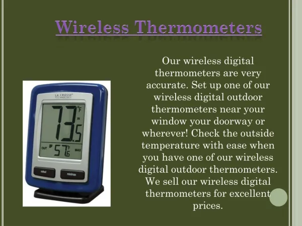
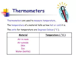
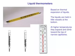
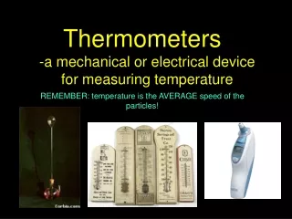
THERMOMETERS. There are many different types of thermometers but they all have one thing in common – they all have a property that changes with temperature. Look at the display of thermometers. How do they work?

THERMOMETERS
E N D
Presentation Transcript
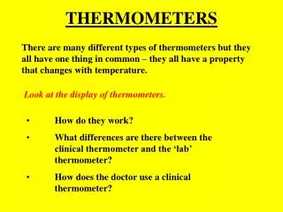
THERMOMETERS There are many different types of thermometers but they all have one thing in common – they all have a property that changes with temperature. Look at the display of thermometers. • How do they work? • What differences are there between the clinical thermometer and the ‘lab’ thermometer? • How does the doctor use a clinical thermometer?
THERMOMETERS -answers • A thermometer must have some physical property that changes with temperature e.g. mercury expands when heated. • A clinical thermometer has a small range, a kink and a triangular shape. • Shake thermometer; place under tongue for 60sec; remove for reading.
SOUND Sounds are produced by vibrations. For a sound to be transmitted, there must be particles between the source and receiver. Sounds can travel through solids, liquids and gases, but not through a vacuum. The sound wave travels at 340ms-1 through the air. They travel fastest through solids and slowest through gases.
SPEED OF SOUND LEARNINGINTENTION To calculate the speed of sound experimentally. APPARATUS fast timer;2 microphones,metre stick METHOD Place the 2 microphones 1m apart. Switch on timer. Clap hands above start microphone. Record time in table. Repeat twice more. Calculate average time using v = d/t. RESULTS Speed of sound equals 340m/s. CONCLUSION
STETHOSCOPE The stethoscope is used in medicine to listen to heart and lung sounds. The large closed bell is used to listen to high frequency lung sounds and the small open bell is used to listen to low frequency heart sounds. The bell picks up sound. The tubing carries the sound to the ears. Earpieces keep out external noises and keep sound loss to a minimum.
HEARING RANGE AND ULTRASOUNDS The human hearing range is between 20Hz and 20000Hz. Frequencies above this are called ULTRASOUNDS and cannot be heard by humans, though animals like bats and dogs can hear them. Ultrasounds are used in medicine to produce scans of the inside of the body (INSIDE WOMB) and high intensity ultrasounds are used to break up kidney stones.
HOW ULTRASOUND SCANNERS WORK. Ultrasonic waves are produced by a device called a transducer. This is pressed against the skin of patient (jelly) and a narrow beam of ultrasounds scan the body. Some of these are reflected back whenever they pass from one type of tissue to another. A computer analyses the information.
SOUND LEVEL AND NOISE POLLUTION Sound level is measured in decibels (dB). Some important sound levels to remember are: threshold of hearing 0dB normal conversation at 1m 60dB danger level 80dBthreshold of pain 140dB Your hearing can be damaged by over exposure to loud noises. Ear protectors are used to absorb the sound energy.
BENDING LIGHT LEARNING INTENTION To show the change of direction of light as it passes from air to glass, then glass to air. APPARATUS Power supply, ray box, single slit, rectangular block. INSTRUCTIONS Normal
RESULTS Normal CONCLUSION The beam of light moves towards the NORMAL when it passes from air to glass and away from the normal when it passes from glass to air.
Refraction of light in a converging lens. LEARNINGINTENTION To investigate refraction of light in 2 converging lenses of different thicknesses. APPARATUS Power supply, ray box, triple slit 2 convex lenses (thin & thick). INSTRUCTIONS (a) Thin convex (b) Thick convex
RESULTS (a) Thin convex (b) Thick convex CONCLUSION The thick convex lense refracts the light more.
Refraction of light in a diverging lens. LEARNINGINTENTION To investigate refraction of light of a diverging lens. APPARATUS Power supply, ray box, triple slit, concave lens. INSTRUCTIONS Concave lense
RESULTS CONCLUSION The concave lens causes the light to spread out.
NORMAL SIGHT Lens Retina In normal sight the image is focused accurately onto the retina when looking at near or far objects.
SHORT SIGHT Lens Retina Concave lens for correction In short sight the image is focused in front of the retina. Distance vision is worse than near vision
LONG SIGHT Lens Retina Convex lens for correction In long sight the image is focused behind the retina. Near vision is worse than distance vision.
FOCAL LENGTH The focal length of a lens is illustrated below. Focal length Watch the demonstration on how to measure the focal length of a converging lens. Now you do the same.
POWER OF A LENS The power of a lens is related to its focal length as shown :- power = 1/focal length focal length = 1/power Power is measured in dioptres(D) and the focal length must be in metres. A convex lens has a positive power and a concave lens has a negative power.
LASER • A laser is a concentrated high- energy beam of light. A carbon dioxide laser is used to treat tumours. An argon laser is used to repair damage to the retina of the eye. An argon laser is also used to remove birthmarks.
ELECTROMAGNETIC SPECTRUM The electromagnetic spectrumVisible light is just one type of electromagnetic radiation. There are various types of electromagnetic radiation with longer wavelengths of light than red light and with shorter wavelengths than violet light. All the different types of electromagnetic waves travel at the same speed through space.
X-RAYS X-rays: pass through soft body tissue but are absorbed by dense bones in the body. X-rays darken an unexposed photographic film. X-rays: are used to detect breaks in bones - the break showing up as a dark line on a photographic film while the bone appears white. CAT scanner: uses x-ray images from a number of angles to build up a 3D image of the inside of the body.
ULTRAVIOLET • Ultravioletradiation is used to treat skin conditions such as acne. UV radiation is also used to sterilise equipment because it can kill harmful bacteria. Too much exposure to uv radiation may produce skin cancer. • Ultraviolet radiation is also used in: • Sun beds Security pens Fluorescent lights (coatings inside the light absorb the ultraviolet light and re-emit it as visible light).
INFRARED • Infraredradiation is another term for heat. Infrared radiation is used to treat strained muscles and tissue. Infrared radiation is also used to diagnose tumours. This works because a tumour emits more infrared radiation than healthy tissue does. This radiation can be detected on a thermogram - a photograph taken using infrared radiation.
RADIATION Radiation can be defined as: energy given off by the nucleus of an atom in the form of particles or rays. Radiation is in every part of our lives. It occurs naturally in the earth and can reach us through cosmic rays from outer space. Radiation may also occur naturally in the water we drink or the soils in our backyard. It even exists in food, building materials, and in our own human bodies. Radiation is used for scientific purposes, medical reasons, and to power some submarines. We can also come into contact with radiation through sources such as X-rays, nuclear power plants, and smoke detectors.
THE ATOM All nuclear radiation comes from inside the atom. An atom consists of protons (+) and neutrons (no charge) surrounded by electrons(-)
ALPHA RADIATION Alpha radiationAlpha radiation consists of alpha particles. An alpha particle is identical to the nucleus of a helium atom, which comprises two protons and two neutrons. Alpha radiation can be stopped by a sheet of paper.
BETA RADIATION Beta radiationBeta radiation consists of high energy electrons emitted from the nucleus. These electrons have not come from the electron shells or energy levels around the nucleus. Instead, they form when a neutron splits into a proton and an electron. The electron then shoots out of the nucleus at high speed. Beta radiation can be stopped by a thin sheet of aluminium.
GAMMA RADIATION Gamma radiationGamma waves have a very high frequency. Gamma radiation cannot be seen or felt. It mostly passes through skin and soft tissue, but some of it is absorbed by cells. • Gamma radiation is used to: • Sterilise surgical instruments. • Kill harmful bacteria in food. • Kill cancer cells (note that lower doses of gamma radiation could cause cells to become cancerous). Gamma radiation can be stopped by dense materials like lead.
IONISATION Nuclear radiation can cause electrons to break free from the atom. When this happens the atom has become positively charged (a positive ion). Alpha radiation causes much greater ionisation than beta or gamma.
Photographic fogging When light falls on photographic film the chemical on the surface changes and the film blackens (fogs). Alpha, beta and gamma radiations have a similar effect on photographic film and can therefore be used to detect it (film badges for people who work in the nuclear industry).
RADIOACTIVE DECAY & HALF LIFE The activity of a radioactive source is a measure of how much radiation it is giving out. The unit of activity is the becquerel (Bq). If a source has an activity of 1Bq then 1 atom disintegrates each second and gives out a particle of radiation. The half life of a source is the time taken for the source to half it’s original value.
MEASURING HALF-LIFE First of all the background count rate is measured using a GM tube connected to a counter. Detecting radiation with a GM Tube The count rate from the source is measured at regular fixed intervals over a period of time. The background count rate is subtracted from each measurement and the actual count rate from the source is measured.
A graph of the count rate of the source against time is plotted. From the graph, the time taken for the count rate to fall by half is measured.
RADIATION DOSE • The radiation dose received by body tissue depends on the type of radiation absorbed by the tissue eg whether the radiation is alpha, beta, gamma or some other type of radiation such as X-rays. The radiation dose received also depends on the energy of the radiation. A way of expressing the radiation dose received from different sources is in terms of a quantity called 'equivalent dose'. Equivalent dose is measured in sieverts (Sv). A dose of one sievert from an alpha radiation source, for example, is equivalent to a dose of one sievert from a beta radiation source or any other source of radiation.