Immunohistochemical Analysis of Rabies and HIV Co-infection in Neuronal Cells
This slide illustrates a rabies-infected neuronal cell exhibiting intracytoplasmic inclusions. The red staining highlights regions containing rabies viral antigens, through the use of immunohistochemistry (IHC) with the avidin-biotin complex (ABC) technique. Additionally, confocal microscopy reveals double immunofluorescent staining for RCA-1 (red) and HIV gp41 protein (green), with evidence of HIV co-infection indicated by the yellow co-localization with RCA-1-stained macrophages. An immunofluorescence assay demonstrates SARS-CoV-infected Vero cells treated with patient serum post-symptom onset.

Immunohistochemical Analysis of Rabies and HIV Co-infection in Neuronal Cells
E N D
Presentation Transcript
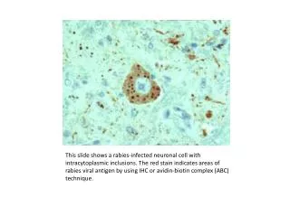
This slide shows a rabies-infected neuronal cell with intracytoplasmic inclusions. The red stain indicates areas of rabies viral antigen by using IHC or avidin-biotin complex (ABC) technique.
Confocal microscopy of double immunofluorescent staining for RCA-1 (red) and HIV gp41 protein (green). At least one of perivascular RCA-1 stained macrophages is infected with HIV, as evidenced by co-localization of RCA-1 and gp41 (yellow).
Immunofluorescence assay (IFA): SARS-CoV-infected Vero cells incubated with patient serum (1:50 dilution) obtained 11 days after the onset of symptoms, showing cytoplasmatic fluorescence. (Source: Institute for Medical Virology, Director: W. Doerr)