Download
1 / 25
250 likes | 390 Vues
The early minutes of reperfusion provide critical opportunities for cardioprotection, yet cardiomyocyte hypercontracture emerges as a major feature of reperfusion injury. This condition, marked by contraction band necrosis, reflects severe hypercontracture and is driven by mechanisms such as Ca²⁺ overload and rigor-type contracture due to ischemia. Various strategies to mitigate hypercontracture include phosphatase inhibition, modulation of Ca²⁺ oscillations, and enhancing mitochondrial energy recovery. Understanding these mechanisms and therapeutic approaches is essential for improving outcomes after myocardial infarction.

E N D
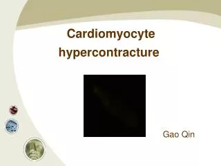
Cardiomyocyte hypercontracture Gao Qin
Background • The first minutes of reperfusion repre-sent a window of opportunity for cardioprotection • Development of cardiomyocyte hyper- contracture is a predominant feature of reperfusion injury
Background • A pattern of contracture and necrotic cell injury “Contraction band necrosis” can be found during the early stage of the infarct • “Contraction band necrosis” reflects hypercontracture of myocytes
Infarct size Contraction band necrosis
Confocal image of adult ventricular myocyte loaded with TMRM
What causes contracture ?
Mechanisms of contracture • Ischemia-induced contracture Rigor-type mechanism • Reperfusion-induced hypercontracture Ca2+ overload-induced contracture Rigor-type contracture
Ischemia-induced contracture • Rigor-type mechanism Low cytosolic ATP myofibrillar shortening cytoskeletal defects cardiomyocytes more fragile and susceptible to mechanical damage
Reperfusion-induced hypercontracture • Much greater myofibrillar shortening and cytoskeletal damage • Aggravated form of contracture lead to a marked rise in end-diastolic pressure
Two causes for reperfusion- induced hypercontracture • Ca2+ overload-induced contracture energy recovery rapid but cytosolic Ca2+ load high • Rigor-type contracture energy recovery very slow
Two causes for reperfusion- induced hypercontracture
NCE forward NCE reverse Ca2+overload-induced hypercontracture NHE NBS NHE:Na+/H+ exchanger NCE:Na+/Ca2+ exchanger NBS: Na+/HCO3- symporter
Ca2+overload-induced hypercontracture • Cyclic uptake and release of Ca2+ by the sarcoplasmic reticulum (SR) in reoxygenated cardiomyocyte (reperfused heart) triggers a Ca2+ oscillations-induced hypercontracture Reperfusion
Ca2+overload-induced hypercontracture
Treatments • Initial ,time-limited inhibition of the contractile machinery • Phosphatase 2,3-butanedione monoxime • cGMP-mediated effectors (NO,ANP) • Cytosolic acidosis (-)NHE,(-)NBS reduce the Ca2+ sensitivity of myofibrils
Treatments • Reducing SR-dependent Ca2+oscillations • Interfering with SR Ca2+ ATPase or SR Ca2+ release • Interfering with SR Ca2+ sequestration • Inhibiting Ca2+ influx
Rigor hypercontracture • Prolonged ischemia mitochondria can not recover cardiomyocytes may contain very low concentrations of ATP at the early of re- oxygenation induce rigor-type contracture major contributor to reoxygenation-induced hypercontracture Ca2+ independent
Treatments • Improving the conditions for energy recovery • application of mitochondrial energy substrates (succinate) Accelerating oxidative energy production • protecting mitochondria from compulsory Ca2+ uptake Resuming oxidative phosphorylation
Spread of hypercontrature gap junctionspreads cell injury
Protective signaling pathways • PKC-dependent signaling • PKG-dependent signaling • PI 3-kinase signaling
PKC- dependent signaling • Not improve the cellular state of energy or the progressive loss of control of cation homeostasis • But attenuate the development of hypercontracture
PI 3-kinase signaling • Insulin protects the cells against hypercontracture through a PI 3-kinase-mediated pathway
References • H.M. Piper, Y. Abdallah, C. Schäfer. The first minutes of reperfusion: a window of opportunity for cardioprotection. Cardiovascular Research 61 (2004) 365– 371 • Y. Ladilov, Ö. Efe, C. Schäfer, H.M. Piper et al. Reoxygenation- induced rigor-type contracture.Journal of Molecular and Cellular Cardiology 35 (2003) 1481–1490 • H. Michael Piper, K. Meuter, C.Schäfer. Cellular mechanisms of ischemia-reperfusion injury.The Annals of Thoracic Surgery 75 (2003) 644–648 • H.M. Piper, D. Garcıa-Dorado, M. Ovize. A fresh look at reperfusion injury.Cardiovascular Research 38 (1998) 291–300