Download
1 / 90
920 likes | 1.18k Vues
Liver and Cardiac Enzymes and Drug Interference. By Zohreh Rahimi Professor of Clinical Biochemistry. TESTS OF LIVER INJURY PLASMA ENZYME LEVELS

E N D
Liver and Cardiac Enzymes and Drug Interference • By • ZohrehRahimi • Professor of Clinical Biochemistry
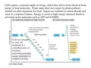
TESTS OF LIVER INJURY • PLASMA ENZYME LEVELS • As metabolically complex cells, hepatocytes contain high levels of a number of enzymes. With liver injury, these enzymes may leak into plasma and can be useful for diagnosis and monitoring of liver injury. • Cellular Locations of Enzymes • Cytoplasmic enzymes include lactate dehydrogenase (LD), aspartate aminotransferase (AST), and alanine aminotransferase (ALT). Mitochondrial enzymes, such as the mitochondrial isoenzyme of AST, are released with mitochondrial damage. Canalicular enzymes, such as alkaline phosphatase and γ-glutamyl transferase (GGT), are increased by obstructive processes.
Mechanisms of Enzyme Release • Enzymes are released from hepatocytes as a result of injury to the cell membrane that directly causes extrusion of the cytosolic contents. • In addition, agents like ethanol cause release of mitochondrial AST from hepatocytes and its expression on cell surfaces. • Accumulation of bile salts with canalicular obstruction causes release of membrane fragments with attached canalicularenzymes.
Increased synthesis of GGT, and to a lesser extent alkaline phosphatase, can occur with medications that induce microsomal enzyme synthesis, notably ethanol, phenytoin, and carbamazepine. • Erythromycinthrough recurrent intrahepatic cholestasis increases alkaline phosphatase and in 15% of patients increases bilirubin by cholestatic effects. Cephalexin through transient hepatitis and cholestatic jaundice increase alkaline phosphatase.
Aminotransferases (Transaminases) • Two diagnostically very useful enzymes in this category are AST or aspartate amino transferase, also known as serum glutamate oxaloacetate transaminase(SGOT), and ALT or alanine amino transferase, formerly called serum glutamate pyruvate transaminase (SGPT). These enzymes catalyze reversibly the transfer of an amino group of AST or ALT to α-ketoglutarate to yield glutamate plus the corresponding ketoacid of the starting amino acid (i.e., oxaloacetate or pyruvate, respectively). Both enzymes require pyridoxal phosphate (vitamin B6) as a cofactor.
AST and ALT have respective blood half-lives of 17 and 47 hours, respectively, and have upper reference range limits of around 40 IU/L. AST is both intramitochondrial and extramitochondrial, but ALT is completely extramitochondrial. AST is ubiquitously distributed in the body tissues, including the heart and muscle, whereas ALT is found primarily in the liver, although significant amounts are also present in the kidney.
Because the serum assays for both ALT and AST required vitamin B6supplied by the patient’s serum, and the patient, an alcoholic, was vitamin B6 deficient (common in alcoholics), the assays for both enzymes showed normal to low levels caused by the absence of vitamin B6. Upon therapeutic intervention, when vitamins were administered, sufficient serum levels of vitamin B6were present to allow full enzyme activities. This clinical history illustrates the central role of pyridoxal phosphate in enzyme catalysis by AST and ALT, and the importance of understanding the chemical basis for enzyme assays.
With most forms of acute hepatocellular injury, such as hepatitis, AST will be higher than ALT initially because of the higher activity of AST in hepatocytes. Within 24 to 48 hours, particularly if ongoing damage occurs, ALT will become higher than AST, based on its longer half-life. An exception to these observations is seen in acute alcohol-induced hepatocyte injury, as in alcoholic hepatitis. Studies suggest that alcohol induces mitochondrial damage, resulting in the release of mitochondrial AST, which, besides being the predominant form of AST in hepatocytes, has a significantly longer half-life than do extramitochondrial AST and ALT. This frequently results in the disproportionate elevation of AST over ALT, yielding an AST/ALT quotient, also called the DeRitis ratio, of 3 to 4:1 in alcohol-induced liver disease.
High AST/ALT ratios suggest advanced alcoholic liver disease. It should also be noted that many alcoholics are vitamin B6 deficient, causing lower rates of synthesis of ALT and suppression of existing ALT activity. • In chronic hepatocyte injury, mainly in cirrhosis, ALT is more commonly elevated than AST; however, as fibrosis progresses, ALT activities typically decline, and the ratio of AST to ALT gradually increases, so by the time cirrhosis is present, AST is often higher than ALT. However, in end-stage cirrhosis, the levels of both enzymes generally are not elevated and may be low as the result of massive tissue destruction.
Overall, ALT activity is more specific for detecting liver disease in nonalcoholic, asymptomatic patients. Mild elevations are often seen in hepatitis C infection. AST is used for monitoring therapy with potentially hepatotoxic drugs; a result more than three times the upper border of normal should signal stopping of therapy.
Assays for AST and ALT • Several variants of assays can be used with these enzymes. In one, alanine for ALT or aspartate for AST is added to force the reaction to the right, yielding glutamate. Production of the latter is then coupled to the enzyme glutamate dehydrogenase, in the so-called indicator reaction, yielding α-ketoglutarate. In this reaction, nicotinamide adenine dinucleotide (NAD) is converted to NADH (reducing agent derived from NAD), which can be measured as an increase in absorbance at 340 nm. • It is vital that pyridoxal phosphate be present in sufficient quantity to allow these reactions to proceed.
Lactate Dehydrogenase • This cytosolic glycolytic enzyme catalyzes the reversible oxidation of lactate to pyruvate. Five major LD isozymes exist, consisting of tetramers of two forms, H and M, the former having high affinity for lactate, the latter for pyruvate. Progressing from HHHH to MMMM, the five possible isozymes are labeled LD1 to LD5. LD1 and LD2 predominate in cardiac muscle, kidney, and erythrocytes. LD4 and LD5 are the major isoenzymes in liver and skeletal muscle. • The upper reference range limit for total LD activity in serum is around 150 IU/L. Serum LD levels become elevated in hepatitis; often, these increases are transient and return to normal by the time of clinical presentation because LD isozymes originating in liver (LD4 and LD5) have relatively low activity in hepatocytes relative to plasma (about 500 times) and a half-life of approximately 4 to 6 hours.
More important is the large increase in total LD to levels of 500 IU/L or more, combined with a significant increase in alkaline phosphatase (ALP)to levels of greater than 250 IU/L, in the absence of other dramatic abnormalities in liver function enzyme levels, especially AST and ALT. These selective increases often accompany space-occupying lesions of the liver, such as metastatic carcinoma and primary hepatocellular carcinoma or, rarely, benign lesions, such as hemangiomata and adenomas. The source of the LD, most often the LD5 isozyme, is not clear because it can originate from hepatocytes, from the tumor, or from both. The rise in ALP is due to blockage of local canaliculi and ductulesby the masses in the liver.
Enzymes Primarily Reflecting CanalicularInjury • These enzymes are located predominantly on the canalicularmembrane of the hepatocyte and include alkaline phosphatase, γ-glutamyl transferase, and 5′-nucleotidase. In contrast to cytoplasmic enzyme activities, canalicular enzyme activities within hepatocytes are typically quite low; focal hepatocyte injury seldom causes significant increases in canalicular enzyme levels. • Alkaline Phosphatase • ALP is present in a number of tissues, including liver, bone, kidney, intestine, and placenta, each of which contains distinct isozymes that can be separated from one another by electrophoresis. Total ALP in serum is mainly present in the unbound form and, to a lesser extent, is complexed with lipoproteins or rarely with Igs.
ALP in the liver, which has a half-life of about 3 days, is a hepatocyticenzyme that is found on the canalicular surface and is therefore a marker for biliary dysfunction. The bone isozyme is particularly heat labile, allowing it to be distinguished from the other major forms. In addition, small intestinal and placental ALP is antigenically distinct from liver, bone, and kidney ALP. The bulk of ALP in the serum of normal individuals is made up of liver.
In obstruction of the biliary tract by stones in the ducts or ductules, or by infectious processes resulting in ascending cholangitis, or by space-occupying lesions, biliary tract ALP rises rapidly to values sometimes in excess of 10 times the upper limit of normal. The reasons for this increase probably include a combination of increased synthesis and decreased excretion of ALP. • A high molecular weight ALP appears in serum in cholestasis. This ALP is attached to fragments of canalicular membrane.
γ-Glutamyl Transferase • This enzyme regulates the transport of amino acids across cell membranes by catalyzing the transfer of a glutamyl group from glutathione to a free amino acid. Its major use is to discriminate the source of elevated ALP (i.e., if ALP is elevated and GGT is correspondingly elevated, then the source of the elevated ALP is most likely the biliary tract). The highest values, often greater than 10 times the upper limit of normal, may be found in chronic cholestasis due to primary biliary cirrhosis or sclerosing cholangitis. This enzyme is also elevated in about 60% to 70% of those who chronically abuse alcohol, with a rough correlation between the amount of alcohol intake and GGT activity.
GGT is often increased in alcoholics even without liver disease; in some obese people; and in the presence of high concentrations of therapeutic drugs, such as acetaminophen and phenytoin and carbamazepine (increased up to five times the reference limits), even in the absence of any apparent liver injury.Also, its level increases in the presence of NSAID (Aspirin, brophen). Similarly, elevated GGT and albuminuria have been found to predict the development of hypertension. • These increases in GGT may occur in order to restore glutathione used in the metabolism of these drugs. Glutathione is conjugated to these drugs via the glutathione S-transferase system, and the complex is then excreted.
Most assays for GGT utilize the substrate γ-glutamyl–p-nitroanilide. In the reaction catalyzed by GGT, p-nitroanilineis liberated and is chromogenic, enabling this colored product to be measured spectrophotometrically. • Other Enzymes • 5′-Nucleotidase activity is increased in cholestatic disorders with virtually no increase in activity in patients with bone disease. Measurement of 5′-nucleotidase can corroborate the elevation of ALP from a hepatic source.
Medication-induced liver injury ranges from very mild to very severe. Virtually any prescription medication has the capacity to cause liver enzymes to rise in a given individual, and not all elevations are persistent or worrisome. • Prescribed medicines most commonly associated with liver injury and elevation of the transaminase enzymes, AST and ALT, include non-steroidal pain relievers, antibiotics, cholesterol-lowering statins, anti seizure medications, and drugs for tuberculosis.
Acetaminophen(Tylenol) is a commonly used prescription pain reliever and an ingredient in a wide variety of products on drug store shelves. While acetaminophen is safe to use at recommended dosages, overdoses may result in liver damage that can unfold over 2 to 3 days. Sometimes, such liver damage is severe enough to be called acute liver failure. The liver enzymes AST and ALT are usually elevated in these cases. It is also important to be aware that liver toxicity from acetaminophen is especially prevalent when patients drink alcohol while using acetaminophen-containing medicines.
Statins • Cholesterol-lowering statins such as atorvastatin (Lipitor) can cause certain liver enzymes levels to spike. It is common for liver enzymes to go up mildly in people taking statins. Although rare, more elderly patients are more likely to have adverse liver reactions to statins as they are at higher risk for organ failure in general. • Statin therapy has been associated with elevated hepatic transaminases in up to 1-3% of patients. • This usually is dose dependent and occurs within the first three months of commencing therapy, and is not usually associated with any long-term hepatic dysfunction.
The possibility of liver intoxication at therapeutic doses of paracetamol (maximal dailydose of 4 x 1 g) is supported by retrospective, but not by controlled prospective studies. Intended or suicidal overdosages are frequently misjudged in retrospective reports. Transient increases in transaminase values (> 3 x upper limits of normal) after regular doses of paracetamol are not proof of hepatic damage unless associated with corresponding symptoms or laboratory changes indicative of compromised hepatic function (total bilirubin, INR). There is insufficient evidence of liver injury by paracetamol at regular dose levels.
Antibiotics • Increases in liver enzymes are a common side effect of many different types of antibiotics such as amoxacillin, ciprofloxacin and erythromycin. However, it is difficult to predict which patients are most likely to have liver problems from antibiotic use, as many patients using antibiotics already have liver enzyme changes due to other conditions. If you are at risk for liver complications or have had liver problems in the past, it may monitor your liver enzyme levels while you are taking antibiotics to make sure no liver complications come up during your treatment.
Anti-epileptic drugs such as carbamazepine and tuberculosis medicines such as Rifampin (Rifadin) are commonly associated with changes in liver enzyme levels and liver function. Other common prescription drugs that can also increase liver enzyme levels include antidepressants and many antiviral drugs. • It is also important to recognize the signs of liver toxicity, including a yellowing of the skin known as jaundice, pain in the abdomen, loss of appetite and nausea.
Drugs that may cause a raised alkaline phosphatase include: • nitrofurantoin • phenytoin • erythromycin • disulfiram
Dipyrone (noramidopyrinemethanesulfonate) is aneffective analgesic, antipyretic, and anti inflammatorydrug. • The minimum concentrations of dipyrone producing interference ranged from22 to 1423 micro mol/L, depending on the serum analytebeing measured. • 2 g of dipyrone administered intravenously has a statistically significant effect on the measurement of CK, LD, uric acid,triglycerides, and cholesterol.
ALT • ALT measurement is not only widely used in detecting the incidence, development, and prognosis of liver disease with obvious clinical symptoms, but also provides reference on screening the overall health status during health check-ups. • Some demographic factors, such as gender and age, might also interfere with the ALT level in the general population. • Physically, the ALT enzyme catalyzes the transfer of amino groups from L-alanine to α-ketoglutarate, and the converted products are L-glutamate and pyruvate . • ALT is mainly aggregated in the cytosol of the hepatocyte. ALT activity in hepatic cells is approximately 3000 times higher than serum ALT activity. When liver injury occurs, ALT is released from injured liver cells and causes a significant elevation in serum ALT activity.
Medications and ALT • A randomized controlled trial (RCT) indicated that the estimated odds ratios (ORs) of ALT elevation in active treatment groups (including acetaminophen, hydromorphone+acetaminophen, morphine+acetaminophen, and oxycodone+acetaminophen) were 2.57-3.08 compared to the placebo group involving 343 healthy participants, even at the recommended dose. Another commonly used • medication, statins, also causes mild ALT elevation. The mechanism underlying statin-associated ALT elevation is still unclear. Some scholars have suggested that the ALT elevation in statin users is attributed to cholesterol reduction in hepatocytes and co-morbid conditions, rather than liver damage or dysfunction.
Coffee consumption and ALT • Of note, coffee intake might be a protective factor against ALT elevation. In some studies, there was a 50% and 70% decrease in ALT elevation amongst participants who consumed >2 cups of coffee/day or ≥373 mg of caffeine, respectively, compared to participants who did not consume coffee. The protective effects of caffeine has been atributed to antioxidant activity. • Liver function tests and diet • There is a clear relationship between the marked rises in transaminases and the number of days on the HCHC (High carbohydrate, high calorie) diet. Such a powerful relationship was not found with the isocaloric HFHC diet and demonstrates the importance of carbohydrate rather than calories as the prime factor in the changes found.
However, some small changes were apparent in ALT when subjects ate the HFHC (high fat, high calorie) diet. • A much higher proportion of the calories in the carbohydrate fraction of the HCHC diet was due to sucrose than in the other diets. In contrast, the amount of starch in each of the high-calorie diets was nearly the same and about double that in the balanced normal calorie diet. This implies that it is the amount of sucrose in the high-carbohydrate diet which mainly underlies the marked rises in transaminases. • It has been suggested previously that a rise in transaminase activity may be due to the fructose moiety of the sucrose in the diet causing damage to hepatocytes or to lipid deposition in the liver.
It seems more plausible that the transaminases are induced by the increased flux of carbohydrate through glycolysis and related pathways. The greater effect upon ALT compared with AST might be explained by the fact that the former enzyme is involved directly with pyruvate metabolism whereas AST is more indirectly related to carbohydrate metabolism. Hepatic enzyme induction by increased availability of substrate, such as when certain drugs, e.g. barbiturates, phenytoin, ethanol, etc. are taken regularly, is a well-recognized phenomenon. • The HCHC diet also produced small but significant rises in ALP and gGT activities.
P450 enzyme classification • In man there are around 30 CYP enzymes which are responsible for drug metabolism and these belong to families 1–4. It has been estimated, however, that 90% of drug oxidation can be attributed to six main enzymes: CYP 1A2, 2C9, 2C19, 2D6, 2E1 and 3A4. The most significant CYP isoenzymes in terms of quantity are CYP3A4 and CYP2D6. • Diet and environment • The addition of food supplements containing cruciferous vegetables, such as cabbage, could increase the activity of both CYP1A1 and CYP1A2 by a factor of 70. A further example relates to induction of CYP2B1 by diallylsulphide in garlic.
Another important cause of morbidity due to enzyme inhibition is citrus fruit. The most important of these is grapefruit juice, which contains a number of potent CYP enzyme inhibitors. These include the plant alkaloids naringin, naringenin and bergamottin. In particular, CYP3A4 is inhibited, leading to altered drug disposition of a number of substances including the antihistamine terfenadine, which can result in fatal cardiac arrhythmia. • Atmospheric pollution is also a cause of enzyme induction.
Alcohol and cigarette smoke • Liver enzyme induction in cigarette smokers is complex, due to the multiplicity of substances which can be detected in cigarettes. The polycyclic aromatic hydrocarbons (PAHs) typically induce CYP1A1 and CYP1A2. • Cigarette smoke also contains a number of small molecules, including various alcohols, styrene, acetone and vinyl chloride, which are also inhaled. These are substrates for CYP2E1 and this enzyme is also induced. Nicotine may also play a part in liver enzyme induction and in animal studies it induces CYP1A, CYP2B6 and CYP2E1. • There is an interesting synergy between alcohol ingestion and cigarette smoking. Although alcohol is primarily metabolized by alcohol dehydrogenase,CYP2E1accounts for around 20% of its breakdown.
The synergistic effects of nicotine and alcohol on the induction of liver CYP2E1 also may explain the higher ethanol elimination rates among smokers and the high percentage of smokers among alcoholics. There may be health implications, therefore, as a result of increased CYP2E1 activity for those patients who are prescribed nicotine, either as nicotine replacement therapy or as part of therapy for illnesses such as ulcerative colitis, Alzheimer’s disease and Parkinson’s disease.
Cordaron(Amiodaron) • Cordaron is administered in ventricular arrhythmia cases or - in severe cases - intravenously. It is known that Cordaron can cause hepatic damage, especially in patients sensitive to the drug.
Theophylline • Theophylline is generally used in departments of respiratory diseases. Theophylline over dosage can cause severerhabdomyolysiswith elevated CK level. It is suggested that the CK activity to be examined only in samples with theophylline concentrations above 30 mg/L. • Acetaminophen • Acetaminophen (Tylenol) is a commonly used prescription pain reliever and an ingredient in a wide variety of products on drug store shelves. While acetaminophen is safe to use at recommended dosages, overdoses may result in liver damage that can unfold over 2 to 3 days. Sometimes, such liver damage is severe enough to be called acute liver failure. Acetaminophen toxicity is the number one cause of acute liver failure in the United States, and the liver enzymes AST and ALT are usually elevated in these cases. It is also important to be aware that liver toxicity from acetaminophen is especially prevalent when patients drink alcohol while using acetaminophen-containing medicines.
Antibiotics • Increases in liver enzymes are a common side effect of many different types of antibiotics such as amoxacillin, ciprofloxacin and erythromycin. However, it is difficult to predict which patients are most likely to have liver problems from antibiotic use, as many patients using antibiotics already have liver enzyme changes due to other conditions. If you are at risk for liver complications or have had liver problems in the past, your doctor may monitor your liver enzyme levels while you are taking antibiotics to make sure no liver complications come up during your treatment.
Diagnosis of acute myocardial infarction • The diagnosis of AMI, as formally established by the World Health Organization (WHO), requires at least two of the following criteria: • A history of chest pain • Evolutionary changes on the ECG • Elevation of serial cardiac enzymes (proteins)
Electrocardiogram One of the mostvaluable contributions of the ECG is in the diagnosis of AMI. • It is usually the first test performed and is often the cornerstone (foundation stone) of the diagnosis. • The initial ECG is diagnostic of AMI in slightly more than 50% of AMI patients. • In about 15% of AMIs, no changes appear on the initial ECG tracing. • Serial tracings over a24-hour period increase its sensitivity to more than 75%. • The ECG changes of an AMI are those of ischemia, injury, and cell death and are reflected by T-wave changes, ST-segment changes, and the appearance of enlarged Q waves, respectively.
Cardiac markers • A cardiac marker is a clinical laboratory test useful in the detection of AMI or minor myocardial injury. • Cardiac markers are most useful when individuals have nondiagnostic ECG tracings. • Individuals with AMI can be categorized into the following four groups. • 1. The first is the group of patientswho present early to the emergency room, within 0 to 4 hours after the onset of chest pain, without diagnostic ECG evidence of AMI. • For laboratory tests to be clinically useful in this group of patients, markers of AMI must be released rapidly from the heart into the circulation. Further, the analytical assays must be sensitive enough to distinguish small changes within the serum reference intervalI.