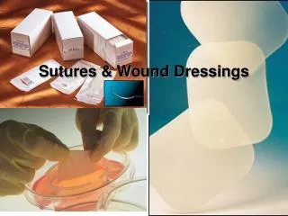
Wound Healing, sutures and needles
170 likes | 774 Vues
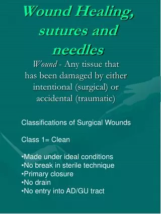
Wound Healing, sutures and needles. Classifications of Surgical Wounds Class 1= Clean Made under ideal conditions No break in sterile technique Primary closure No drain No entry into AD/GU tract.

Wound Healing, sutures and needles
E N D
Presentation Transcript
Wound Healing, sutures and needles • Classifications of Surgical Wounds • Class 1= Clean • Made under ideal conditions • No break in sterile technique • Primary closure • No drain • No entry into AD/GU tract Wound - Any tissue that has been damaged by either intentional (surgical) or accidental (traumatic)
Class 2= Clean contaminated • Primary closure • Wound drained • Minor break in sterile technique • Controlled entry into AD/GU tract • Class 3= Contaminated • Open traumatic wound • Major break in aseptic technique • Acute Inflammation present • Entry into AD/GU tract with spillage • Class 4= Dirty/Infected • Open traumatic wound • Microbial contamination prior to procedure • Perforated viscus • This information needs to be documented onto • the patients chart. The circulating nurse will • the anesthesiologist for this information.
Traumatic Wound • Closed - Skin remains intact/underlying tissue • damaged. • Open wound - Integrity of skin destroyed. • Simple wound - Skin is destroyed, no loss or • destruction of tissue, no foreign • body. • Complicated wound - Tissue lost or destroyed • foreign body may be • present. • Clean wound - Wound edge can be • approximated expected to heal • by first intention. • Contaminated - A dirty object damages integrity • of skin, acute infection, may • need debridement.
Mechanism of injury • Abrasion • Contusion • Laceration • Puncture • Thermal • Chronic Wounds – Persist for an extended • period of time, due to the • physical condition of the Pt. • Inflammatory Process – Protective response to • injury or tissue destruct. • -Classic local signs • Pain • Heat • Redness • Swelling • Loss of function • -When the injured tissue releases histamine • from it’s damaged cells, the histamine causes • the small blood vessels in the area to dilate • Resulting in the above.
Phases of Wound Healing • First Intention (primary union) • Lag/Inflammatory response • Proliferation • Maturation/differentiation • Healing occurs from side to side Lag phase – Begins within minutes of the injury last appox. 3-5 days. This stage controls bleeding through platelet aggregation, delivering blood to the injured site. Proliferation – Begins 3rd post-operative day, continues up to 20 days fibroblasts multiply and bridge wound edges. Maturation/differentiation – Begins on the 14th day post-op lasts until wound is comp- letely healed. This involves inner weaving of collagen fibers.
-Second Intention (granulation) • Wound fails to heal by primary union • Large wounds cannot be approximated • Infection causes breakdown of sutured wound • Dr’s will sometimes allow this following the • removal necrotic tissue or large/wide • debridement's • Heals from inner layer to outside surface • -Third Intention • Two granulated surfaces are approximated • Debrided and purposely left open to heal by • second intention (4-6 days) • Use of antibiotics for pt/special wound care • techniques utilized • Infection free wound is then closed and allowed • to heal by first intention • Works well for contaminated or dirty wounds • (trauma/tissue loss)
Factors Influencing Wound Healing • ( Three main factors) • Age (peds, geriatric) • Nutrition • Disease • Smoking (vasocontricts) • Radiation ( decreases blood supply) • Immune deficiency • Intra-op tissue handling: • Dissection techniques • Duration of surgery • Amt of tissue handling • Secure wound closure • Aseptic techniques • Any microbial contamination of wound could • lead to infection.
Complications of Wound Healing • Dehiscence • Partial layer/ layers of tissue after closure • Mostly seen in debilitated pt’s with friable • tissue. • Abdominal distention • Inappropriate suture material • Improper suture technique • Tearing sensation by pt. • Evisceration – Exposed (protrusion) of the • viscera through the edges of a • totally separated wound. • (emergency situation) • Adhesion – Abnormal attachment of two • surfaces or structures that are • normally separate. • Previous surgeries • Infection • Improper handling • Presence of foreign body (lint, powder • granule)
Intra-operative Wound Care • Local and systemic features • Sterile technique • Tissue perfusion • Antibiotic therapy • Post-operative Wound Care • Drains • Protection of wound with different kinds • of dressings suture 20th Century – Most suture material was made From the intestinal walls of sheep(catgut). Some Preferred silk linen, kangaroo tendon. Catgut – -sold in a dehydrated state -sterilized in open glass tubing (heat) -Sterilized preservative was added and tube was sealed.
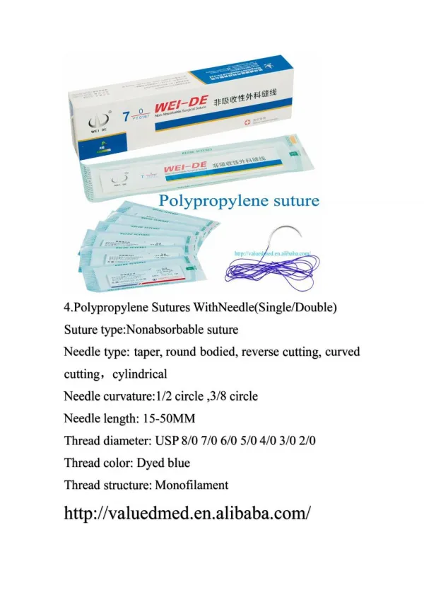
1909- Davis and Geck company perfected a • method for sterilizing the surgical gut. • 1930- D & G introduced specific suture for • Specialties including surgical gut, silk, linen, • Cotton, and celluloid-linen • Types • Absorbable – absorbed by tissue • Nonabsorbable – resists enzymatic digestion • Monofilament – Made of single thread-like • structure • Multifilament– multi threadlike structures braided • twisted into single strand • Suture ties – tying of vessels or tissue with or • with out a needle attached. • Size: • Selected to correspond to type of tissue • Heavy tissue, heavy gauged suture • Gauge is used for sizing method • 11-0 to # 6
characteristics of common suture • Knot strength • Memory • Wicking • Tissue reaction • Absorption rate • Size • Tensile strength • Elasticity examples of suture types • Plain and chromic gut • PDS • Monocryl • Prolene/surgilene • Ethilon/dermalon • Steel • Novafil • Dermalene • Dexon • Silk • Nurolon/Surgilon • Mersilene • Ethibond/Dacron • Pronova • Vicryl
Layer Closure for Abdominal Wounds • Peritoneum • Fascia • Muscle • Subcutaneous • Subcuticular • Skin Needles (Characteristics) • Eye- closed, french eye, swagged on • Point- cutting, rev cut, trocar points • Body-1/2, 3/8 etc • Shape • Needle Point • Cutting Tapered • -Tough tissue -Delicate tissue • -Reverse • -Side; Ophthalmology Blunt • - Kidney, Liver