Download
1 / 18
390 likes | 2.08k Vues
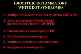
IDIOPATHIC INFLAMMATORY WHITE DOT SYNDROMES. 1. Multiple evanescent white dot syndrome (MEWDS). 2. Acute posterior multifocal placoid pigment epitheliopathy (APMPPE). 3. Punctate inner choroidopathy (PIC). 4. Birdshot retinochoroidopathy.

E N D
IDIOPATHIC INFLAMMATORY WHITE DOT SYNDROMES 1. Multiple evanescent white dot syndrome (MEWDS) 2. Acute posterior multifocal placoid pigment epitheliopathy (APMPPE) 3. Punctate inner choroidopathy (PIC) 4. Birdshot retinochoroidopathy 5. Multifocal choroiditis with panuveitis 6. Serpiginous choroidopathy
MEWDS • Young adults (F > M) • Unilateral • Small, subtle, deep grey-white dots • Orange macular granularity • Mild vitritis Posterior pole Mid-periphery
FA of MEWDS Many hyperfluorescent spots at posterior pole Increase in hyperfluorescence and late leakage from disc
MEWDS • Treatment - nil • Course - 6 weeks • Complications - nil • Prognosis - excellent Residual blind spot enlargement
APMPPE • Young adults (F=M) • Associated with HLA-B7 and DR2 • Bilateral and symmetrical • Large, deep, grey-white, placoid lesions • Mainly at posterior pole • Mild vitritis
FA of APMPPE Early dense hypofluorescence Late staining
APMPEE • Treatment - nil • Course - 4 weeks • Complications - nil • Prognosis - good Residual RPE changes
Punctate inner choroidopathy ( PIC ) • Young, myopic females • Eventually bilateral but asymmetrical • Deep, small, indistinct yellow spots at • posterior pole • All same age • No vitritis
FA of punctate inner choroidopathy Focal hyperfluorescent spots No increase in size
Punctate inner choroidopathy • Treatment - not beneficial • Course - several weeks • Complications - occasional choroidal neovascularization • (CNV ) • Prognosis - guarded Residual dumb-bell shaped scars
FA of CNV in punctate inner choroidopathy Early hyperfluorescence Progressive leakage
Birdshot retinochoroidopathy • Middle-aged (F > M) • HLA - A29 • Deep, oval, creamy, indistinct spots • Radiate from disc towards equator • Moderate vitritis
FA of birdshot retinochoroidopathy • Venous hyperfluorescence • Extensive late intraretinal and disc leakage
Birdshot retinochoroidopathy • Treatment - steroids and immunosuppressive agents • Course - chronic-remittent • Complications - CMO • Prognosis - guarded Residual punched-out, non-pigmented scars
Multifocal choroiditis with panuveitis • Adults (20-50) ( F M ) • Bilateral but asymmetrical • Deep, discrete, grey-yellow spots • Mixed fresh and old • Mid-periphery and fewer at posterior pole • Moderate to severe panuveitis
Multifocal choroiditis with panuveitis • Treatment - steroids • Course - chronic-remittent • Complications - CMO and occasional subretinal fibrosis • Prognosis - guarded Residual pigmented scars
Serpiginous choroidopathy • Middle age (F = M) • Eventually bilateral but asymmetrical • Deep, grey-white lesions with hazy • borders • Initially peripapillary and at • posterior pole then outward spread • Mild vitritis
Serpiginous choroidopathy • Treatment - steroids and immunosuppressive agents • Course - chronic-remittent • Complications - macular scarring • Prognosis - poor Residual scalloped, punched-out areas