Download
1 / 5
50 likes | 175 Vues
Cervical Lymph Nodes. Left S ubclavian Vein. Right S ubclavian Vein. Axillary Lymph Nodes. Left Pleural Cavity (the space) and Parietal Pleura (blue). Transverse Mesocolon. Greater Omentum. Pharyngeal Tonsil (adenoids). Palatine Tonsils. Lingual Tonsil. Pericardial Cavity

E N D
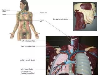
Cervical Lymph Nodes Left Subclavian Vein Right Subclavian Vein Axillary Lymph Nodes Left Pleural Cavity (the space) and Parietal Pleura (blue)
Transverse Mesocolon Greater Omentum Pharyngeal Tonsil (adenoids) Palatine Tonsils Lingual Tonsil
Pericardial Cavity (the space for the heart within the mediastimun) Visceral Pericardium (yellow) Falciform Ligament “Name the inner layer serosa covering this organ” - It is the Visceral Pleura Parietal Pericardium (blue)
Thoracic Cavity (above diaphragm) Abdominal Cavity Greater Omentum Abdominopelvic Cavity Pelvic Cavity (sacral promontory to symphysis pubis) Inguinal Lymph Nodes
9 Regions of the Abdomen: Right Hypochondriac Region Epigastric Region Left Hypochondriac Region Right Lumbar Region Umbilical Region Left Lumbar Region Right Iliac Region Hypogastric Region Left Iliac Region 1 3 2 4 5 6 7 8 9