Download
1 / 21
330 likes | 1.01k Vues
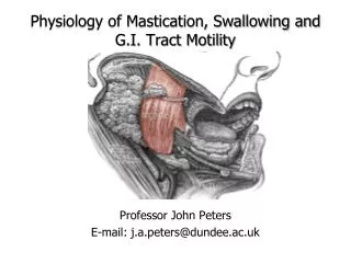
Physiology of Mastication, Swallowing and G.I. Tract Motility. Professor John Peters E-mail: j.a.peters@dundee.ac.uk . Learning Objectives. Following this lecture, students should be able to: List the major functions associated with the mouth

E N D
Physiology of Mastication, Swallowing and G.I. Tract Motility Professor John Peters E-mail: j.a.peters@dundee.ac.uk
Learning Objectives Following this lecture, students should be able to: • List the major functions associated with the mouth • List the 3 main pairs of salivary glands and the nature of their secretions • Describe the concept of primary secretion and its modification by ductal cells • State the functions of saliva • Describe the control(s) of salivary secretion • Describe swallowing in terms of its 'two' phases • Discuss peristalsis in the oesophagus • Discuss the function of the lower oesophagealsphincter • Outline the functions of the stomach
The Basic Digestive Processes (1) • Motility • Mechanical activity mostly involving smooth muscle (skeletal at mouth, pharynx, upper oesophagus and external anal sphincter) • Propulsive movements • Mixing movements • Tonic contractions • Secretion • Into the lumen of the digestive tract occurs from digestive tract and accessory structures in response to hormonal and neural signals. Required for: (i) digestion (ii) protection • Digestive secretions contain: • Water (large volume extracted from plasma) • Electrolytes • Organic compounds (enzymes, bile salts, mucus etc.)
The Basic Digestive Processes (2) • Digestion • Biochemical breakdown (enzymatic hydrolysis) of complex foodstuffs to smaller, absorbable, units • Carbohydrates [mostly polysaccharides (e.g. starch, glycogen) and some disaccharides (e.g. sucrose, lactose)]: converted to monosaccharides (mainly glucose, some galactose and fructose) • Proteins: converted to constituent amino acids • Fats [mostly triglycerides]: converted to monoglycerides and free fatty acids • Absorption • Transfer of the absorbable products of digestion (with water, electrolytes and vitamins) from the digestive tract to the blood or lymph
Structure of the Digestive Tract Wall Mucosa comprises: epithelial cells (absorption) exocrine cells (secrete digestive juices) endocrine gland cells (secrete hormones) lamina propria muscularis mucosa Submucosa contains: connective tissue blood and lymph vessels nerve network – submucous plexus Muscularisexterna comprises: circular muscle nerve network – myenteric plexus longitudinal muscle Serosa contains: connective tissue
The Mouth • Palate • Separates mouth from nasal passages: allows breathing and chewing simultaneously • Uvula • Helps seal off nasal passages during swallowing • Tongue • Guides food, important in speech, and swallowing; major location of taste buds • Pharynx • Common passageway for the respiratory and digestive systems, tonsils on side walls are lymphoid tissues • Lips • Procurement/containment of food • Speech • Teeth • Chewing (mastication), mediated by the masseteric and diagastric reflexes: (i) breaks down food and mixes it with saliva, (ii) stimulates taste buds and (iii) by reflexes following taste bud stimulation increases salivary, gastric, pancreatic and bile secretion
Salivary Secretion and Function • Saliva is secreted by three major pairs of salivary glands: parotid, submandibular and sublingual Functions of Saliva • Parotid - below ear and over the masseter • 25% of total • Lubrication(salivary water and mucus) mouth and food – important for speech and swallowing) • Submandibular - under lower edge of mandible • 70% of total • Solvent(important for taste) • Sublingual in floor of mouth under tongue • 5% of total • Antibacterial(contains lysozyme, lactoferrin and immunoglobulins – important in preventing dental carries ) • Digestion of complex carbohydrate(contains amylase) • Neutralization of acid(contains bicarbonate) • Facilitates sucking by infants(fluid seal)
Formation of Saliva (1) • Occurs in two stages – primary secretion (by the acinus) – secondary modification (by duct cells) • Multiple transporters and ion channels participate in the formation of saliva (which requires ATP) – exact details may differ between glands – only general principles are illustrated schematicallybelow Na+ Cl- Na+, K+, Cl-, HCO3-, H2O H2O HCO3- K+ Duct cells Cells modify secretion by removing Na+ and Cl- and to a lesser extent adding K+ and HCO3-, no movement of H20 – hence diluting Acinus Cells produce a primary secretion with Na+, K+, Cl- and HCO3- content similar to plasma
Formation of Saliva (2) Rate • 0.05 ml/min – sleep • 0.5 ml/min – resting, awake state • 5 ml/min – “actively” salivating Composition • varies according to flow rate • NaCl content much lower than plasma (detection of salty taste?) • HCO3- content: high flow rate ; low flow rate • no glucose (detection of sweet taste?) Reflex regulation (neuronal control) Rate of formation of saliva is increased by: Simple (unconditioned) reflex Conditioned (acquired) reflex (Pavlov’s dog)
Control of Salivary Secretion by Reflexes Simple (unconditioned) Chemo / pressure receptors in mouth activated in presence of food (or other stimulus) Impulses sent via afferent nerves Salivary centre in medulla Acquired (conditioned) Think about, smell, see, hear preparation of appetising dish Cerebral cortex Impulses via extrinsic autonomic nerves - Both sympathetic & parasympathetic stimulation Salivary glands increase production
Role of the Autonomic Nerves in Saliva Secretion Glossopharyngeal and facial nerves Parasympathetic stimulation (Dominant role in ”normal” saliva production) Large volume Watery Enzyme rich (mediated by M3 muscarinic acetylcholine receptors) Note: muscarinic receptor antagonists (e.g. atropine) and antidepressants that block muscarinic receptors cause a dry mouth Postganglionic fibres from superior cervical ganglia Sympathetic stimulation (Dominant at stressful times- dry mouth when nervous?) Small volume Thick Mucus rich (mediated by 1-adrenoceptors)
Swallowing (Deglutition) • Movement of food from the mouth to the stomach occurs in two phases • 1. Oropharyngeal stage (mouth pharynx oesophagus (1 second) Bolus of food must be directed to the oesophagus and away from the nasal passages and trachea – requires a series of highly co-ordinated responses Tongue forces bolus into pharynx at rear of mouth Afferent impulse to swallowing centre in medulla Pressure stimulates pharyngeal pressure receptors Involuntary Voluntary Efferents initiate all-or-nothing reflexsequence of muscle movements (see next slide) Bolus formed in mouth by action of teeth and tongue Food passes through pharynx into oesophagus Upper oesophageal sphincter opens
Oesophageal stage [oesophagus stomach (4-10 seconds)] • Swallowing centre (medulla oblongata) triggers primaryperistaltic wave and closure of the upper oesophagealsphicter • Wave mediated by skeletal muscle in upper oesophagus and smooth muscle in distal regions • Peristalisco-ordinated by the enteric nervous system (cholinergic neurones) • Circular fibres behind bolus squeeze bolus down towards stomach • Longitudinal fibres in front of bolus shorten distance of travel • Lower oesophageal sphincter opens within 2-3 s of the initiation of a swallow (closes after passage of bolus to prevent reflux) • Sticky food may become lodged stimulating local pressure receptors that cause: • secondaryperistaltic wave - more forceful than primary – locally triggered • increased saliva production
Absorption and Secretion by the Oesophagus • Secretion is entirely mucus • Lubricates for passage of food • Protects epithelium from attack by acid and enzymes in gastric juice. Epitheilium itself (stratified squamous) has a rapid turnover • Transit of a bolus of food is far too rapid to permit absorption
The Stomach • J – shaped bag: 50 >1000 ml capacity: relaxes receptively (driven by vagus) to accommodate foodfrom oesophagus • Starting point for digestion of proteins (pepsin and HCl) • Mixes food with gastric secretions to produce chyme • Limited amount of absorption • Stores food before passing it into small intestine as chyme for further digestion and absorption • Secretes approximately 2 litre/day of gastric juice from gastric pits in the gastric mucosa
Gastric Motility • Pacemaker cells (interstitial cells of Cajal) in the fundus establish a basal electrical rhythm (BER) that spreads over the stomach from fundus towards the pyloric sphincter • The BER occurs continuously (3 per minute) and may, or not, generate smooth muscle contraction, dependent upon the excitability of the latter Excitatory stimulus Smooth muscle action potentials Membrane potential (mV) Threshold BER Force (g) Time (s)
Gastric Emptying and Mixing Emptying Mixing Oesophagus Direction of movement of peristaltic contraction Lower oesophageal sphincter Duodenum Pyloric sphincter Peristaltic contraction Peristaltic contraction = movement of chyme
Stomach emptying • Strength of antral wave determines escape of chyme through pyloric sphincter • Governed by: • Gastric factors • Duodenal factors Gastric factors • Rate of emptying proportional to volume of chyme in stomach • Distension increases motility due to stretch of smooth muscle, stimulation of intrinsic nerve plexuses, increased vagusnerve activity and gastrin increase • Consistency of chyme • Emptying facilitated by finely divided, thick liquid chyme
Duodenal Factors • Duodenum must be ready to receive chyme and can delay emptying by: • Neuronal response – the enterogastric reflex – decreases antralperistalic activity through signals from intrinsic nerve plexuses and the autonomic nervous system • Hormonal response – release of enterogastrones [e.g. secretin and cholecystokinin CCK)] from duodenum inhibits stomach contraction Stimuli within the duodenum that drive the neuronal and hormone responses include: • Fat–particularly potent – delay in gastric emptying required for digestion and absorption in small intestine • Acid – time is required for neutralization by bicarbonate secreted from the pancreas – important for optimal function of pancreatic digestive enzymes • Hypertonicity – products of carbohydrate and protein digestion are osmotically active and draw water into the small intestine – danger of reduced plasma volume and circulatory disturbances (e.g. ‘dumping syndrome’) • Distension