Download
1 / 1
10 likes | 93 Vues
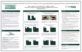
*. *. *. *. *. 538.11 Increased Androgen Receptor Expression in Skeletal Muscle Fibers Leads to Muscle Pathology. Jamie A. Johansen 1 , D. Ashley Monks 3 , S. Marc Breedlove 1,2 , Cynthia L. Jordan 1,2 1 Neuroscience Program, 2 Department of Psychology

E N D
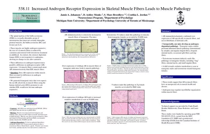
* * * * * 538.11 Increased Androgen Receptor Expression in Skeletal Muscle Fibers Leads to Muscle Pathology Jamie A. Johansen 1, D. Ashley Monks 3, S. Marc Breedlove 1,2, Cynthia L. Jordan 1,2 1Neuroscience Program, 2Department of Psychology Michigan State University; 3Department of Psychology University of Toronto at Mississauga Introduction Results Summary AR immunoreactivity is selectively increased in muscle fibers of transgenic (Tg) mice. Testosterone (T) induces male-like pathology in muscles from transgenic females as revealed by NADH stain. • The spinal nucleus of the bulbocavernosus (SNB) is a sexually dimorphic group of motoneurons that innervate sexually dimorphic perineal muscles, the bulbocavernosus (BC) and levator ani (LA). • These muscles are highly androgen responsive; the size of LA muscle fibers is reduced by castration and restored with testosterone treatment in adulthood. In contrast, the extensor digitorum longus (EDL) is not responsive to androgens, showing no change in size after castration. • These differences in androgen responsiveness parallel a difference in androgen receptor (AR) expression. LA muscle fibers contain much more AR immunoreactivity than EDL muscle fibers. • Question: Does AR expression within muscle fibers account for differences in androgen responsiveness? • We generated transgenic mice that over-express the rat AR selectively in muscle fibers using the human skeletal actin (HSA) promoter to ask whether EDL would now become androgen responsive. • AR immunohistochemistry confirmed over-expression of AR specifically in muscle fibers, and not in fibroblasts or Schwann cells. • Unexpectedly, our mice develop an androgen-dependent pathology. Transgenic males exhibit profound abnormal muscle pathology demonstrated by atrophic and angular-shaped fibers, frequent internal nuclei, occasional split fibers and ring fibers. • Testosterone treatment induced a male-like pathology in transgenic females, including “ring” fibers, internal nuclei, and small angular fibers. • A shift towards oxidative metabolism is seen in transgenic males and testosterone treated transgenic females. Figure 1. Note that AR immunoreactivity is increased only in fibers and not fibroblasts of muscles from transgenic males (right). Internal nuclei, an indication of muscle pathology, also stain positively for AR (arrow). Over-expression of wildtype AR in muscle fibers of transgenic male mice leads to muscle pathology. Figure 4. NADH staining reveals that T treatment has marked effects on EDL muscles from Tg females including alterations in the internal architecture of fibers, and size and shape of the fibers. Note that ring fibers (arrows) are prevalent in the T-treated Tg muscle, and absent in blank-treated Tg muscle. The somewhat darker staining in T-treated female muscles also suggests that T induces a switch to a more oxidative metabolism reminiscent of Tg male mice. Significance • These results suggest that AR in muscle fibers have an unexpected role in muscle health and disease. • Androgens may regulate myofibrillar organization within muscle fibers. T induces male-like pathology in Tg female muscles as revealed by H&E stain. Figure 2. H&E staining of EDL muscles of wildtype (Wt) and transgenic male mice indicate that transgenic muscles contain both abnormally small, angular fibers (black arrow) and hypertrophied fibers (white arrow). Both fiber types contain internal nuclei (stars). Methods Over-expression of wildtype AR leads to increased oxidative metabolism as seen in NADH stain. Acknowledgments Technical support was provided by Cindy Knaff, Heather Malinowski, Diane Redenius, and Laura Baum. These studies were funded by operating grant NIH NS-045195 (CLJ), a grant from the MSU foundation (CJ, SMB) and a postdoctoral fellowship (DAM) from the Canadian Institutes of Health Research (CIHR). • Construct: AR cDNA was subcloned frompCMVAR. cDNAs were ligated into the NotI site of pBSX-HSA. • Histology: Muscles were dissected from transgenic and wildtype males, placed in OCT filled cryomolds and flash frozen in liquid nitrogen. Muscles were then cryostat sectioned at 10um, and stained for Hematoxylin and Eosin (H&E), nicotinamide adenine dinucleotide (NADH) and AR. • Androgen Dependence: To look at androgen dependence, transgenic females were ovariectomized and given either a testosterone filled or blank Silastic capsule. Females were treated for 8 weeks, and then muscles were harvested in a similar way. Figure 3. EDL muscle from wildtype male mice stain moderately for NADH (left). In contrast, the EDL from adult transgenic male mice show increased NADH staining, indicating a switch to a more oxidative metabolism. “Ring” fibers (arrow) are also evident in EDL transgenic muscles stained for NADH. Figure 5. H&E staining in EDL muscles of wildtype and transgenic female mice. Note that fibers in EDL muscles of blank-treated tg females are comparable in size and shape to EDL fibers of both blank- and T-treated wt muscles. In contrast, muscles of tg females exposed to T contain abnormal fibers that exhibit internal nuclei (stars) and “ring” fibers (arrow).