Download

1 / 53
530 likes | 695 Vues
Lesson Overview. 12.1 Identifying the Substance of Genes. Bacterial Transformation. What clues did bacterial transformation yield about the gene?

E N D
Lesson Overview 12.1 Identifying the Substance of Genes
Bacterial Transformation • What clues did bacterial transformation yield about the gene? • By observing bacterial transformation, Avery and other scientists discovered that the nucleic acid DNA stores and transmits genetic information from one generation of bacteria to the next.
Bacterial Transformation • To truly understand genetics, scientists realized they had to discover the chemical nature of the gene. • If the molecule that carries genetic information could be identified, it might be possible to understand how genes control the inherited characteristics of living things. • The discovery of the chemical nature of the gene began in 1928 with British scientist Frederick Griffith, who was trying to figure out how certain types of bacteria produce pneumonia.
Griffith’s Experiments • Griffith isolated two different strains of the same bacterial species. • Both strains grew very well in culture plates in Griffith’s lab, but only one of the strains caused pneumonia. • The disease-causing bacteria (S strain) grew into smooth colonies on culture plates, whereas the harmless bacteria (R strain) produced colonies with rough edges.
Griffith’s Experiments • When Griffith injected mice with disease-causing bacteria, the mice developed pneumonia and died. • When he injected mice with harmless bacteria, the mice stayed healthy. • Perhaps the S-strain bacteria produced a toxin that made the mice sick? To find out, Griffith ran a series of experiments.
Griffith’s Experiments • First, Griffith took a culture of the S strain, heated the cells to kill them, and then injected the heat-killed bacteria into laboratory mice. • The mice survived, suggesting that the cause of pneumonia was not a toxin from these disease-causing bacteria.
Griffith’s Experiments In Griffith’s next experiment, he mixed the heat-killed, S-strain bacteria with live, harmless bacteria from the R strain and injected the mixture into laboratory mice. The injected mice developed pneumonia, and many died. • The lungs of these mice were filled with the disease-causing bacteria. How could that happen if the S strain cells were dead?
Transformation • Griffith reasoned that some chemical factor that could change harmless bacteria into disease-causing bacteria was transferred from the heat-killed cells of the S strain into the live cells of the R strain. • He called this process transformation, because one type of bacteria had been changed permanently into another.
Transformation • Because the ability to cause disease was inherited by the offspring of the transformed bacteria, Griffith concluded that the transforming factor had to be a gene.
The Molecular Cause of Transformation • A group of scientists at the Rockefeller Institute in New York, led by the Canadian biologist Oswald Avery, wanted to determine which molecule in the heat-killed bacteria was most important for transformation.
The Molecular Cause of Transformation • Avery and his team extracted a mixture of various molecules from the heat-killed bacteria and treated this mixture with enzymes that destroyed proteins, lipids, carbohydrates, and some other molecules, including the nucleic acid RNA. • Transformation still occurred. • Avery’s team repeated the experiment using enzymes that would break down DNA. • When they destroyed the DNA in the mixture, transformation did not occur. • Therefore, DNA was the transforming factor.
Bacterial Viruses • What role did bacterial viruses play in identifying genetic material? • Hershey and Chase’s experiment with bacteriophages confirmed Avery’s results, convincing many scientists that DNA was the genetic material found in genes—not just in viruses and bacteria, but in all living cells.
Bacterial Viruses • Several different scientists repeated Avery’s experiments. Alfred Hershey and Martha Chase performed the most important of the experiments relating to Avery’s discovery. • Hershey and Chase studied viruses—nonliving particles that can infect living cells.
Bacteriophages • The kind of virus that infects bacteria is known as a bacteriophage, which means “bacteria eater.” • A typical bacteriophage is shown. • When a bacteriophage enters a bacterium, • it attaches to the surface of the bacterial • cell and injects its genetic information into it. • The viral genes act to produce many new • bacteriophages, which gradually destroy • the bacterium. • When the cell splits open, hundreds of new viruses burst out.
The Hershey-Chase Experiment American scientists Alfred Hershey and Martha Chase studied a bacteriophage that was composed of a DNA core and a protein coat. They wanted to determine which part of the virus—the protein coat or the DNA core—entered the bacterial cell. Their results would either support or disprove Avery’s finding that genes were made of DNA.
The Hershey-Chase Experiment • Hershey and Chase grew viruses in cultures containing radioactive isotopes of phosphorus-32 (P-32) sulfur-35 (S-35) • Since proteins contain almost no phosphorus and DNA contains no sulfur, these radioactive substances could be used as markers, enabling the scientists to tell which molecules actually entered the bacteria and carried the genetic information of the virus.
The Hershey-Chase Experiment • If they found radioactivity from S-35 in the bacteria, it would mean that the virus’s protein coat had been injected into the bacteria. • If they found P-32 then the DNA core had been injected.
The Hershey-Chase Experiment • The two scientists mixed the marked viruses with bacterial cells, waited a few minutes for the viruses to inject their genetic material, and then tested the bacteria for radioactivity. • Nearly all the radioactivity in the bacteria was from phosphorus P-32 , the marker found in DNA. • Hershey and Chase concluded that the genetic material of the bacteriophage was DNA, not protein.
The Role of DNA • What is the role of DNA in heredity? • The DNA that makes up genes must be capable of storing, copying, and transmitting the genetic information in a cell.
The Role of DNA • The DNA that makes up genes must be capable of storing, copying, and transmitting the genetic information in a cell. • These three functions are analogous to the way in which you might share a treasured book, as pictured in the figure.
Storing Information • The foremost job of DNA, as the molecule of heredity, is to store information. • Genes control patterns of development, which means that the instructions that cause a single cell to develop into an oak tree, a sea urchin, or a dog must somehow be written into the DNA of each of these organisms.
Copying Information • Before a cell divides, it must make a complete copy of every one of its genes, similar to the way that a book is copied. • To many scientists, the most puzzling aspect of DNA was how it could be copied. • Once the structure of the DNA molecule was discovered, a copying mechanism for the genetic material was soon put forward.
Transmitting Information • When a cell divides, each daughter cell must receive a complete copy of the genetic information. • Careful sorting is especially important during the formation of reproductive cells in meiosis. • The loss of any DNA during meiosis might mean a loss of valuable genetic information from one generation to the next.
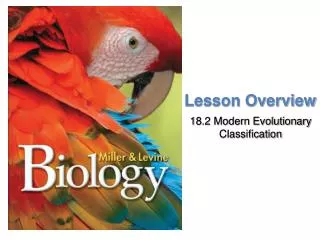
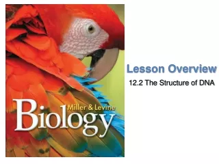
Lesson Overview 12.2 The Structure of DNA
The Components of DNA • What are the chemical components of DNA? • DNA is a nucleic acid made up of nucleotides joined into long strands or chains by covalent bonds.
Nucleic Acids and Nucleotides • Nucleic acids are long, slightly acidic molecules originally identified in cell nuclei. • Nucleic acids are made up of nucleotides, linked together to form long chains. • DNA’s nucleotides are made up of three basic components: a 5-carbon sugar called deoxyribose, a phosphate group, and a nitrogenous base.
Nitrogenous Bases and Covalent Bonds • The nucleotides in a strand of DNA are joined by covalent bonds formed between their sugar and phosphate groups. • DNA has four kinds of nitrogenous bases: adenine (A), guanine (G), cytosine (C), and thymine (T). • The nitrogenous bases stick out sideways from the nucleotide chain. • The nucleotides can be joined together in any order, meaning that any sequence of bases is possible.
Solving the Structure of DNA • What clues helped scientists solve the structure of DNA? • The clues in Franklin’s X-ray pattern enabled Watson and Crick to build a model that explained the specific structure and properties of DNA.
Chargaff’s Rules • Erwin Chargaff discovered that the percentages of adenine [A] and thymine [T] bases are almost equal in any sample of DNA. • The same thing is true for the other two nucleotides, guanine [G] and cytosine [C]. • The observation that [A] = [T] and [G] = [C] became known as one of “Chargaff’s rules.”
Franklin’s X-Rays • In the 1950s, British scientist Rosalind Franklin used a technique called X-ray diffraction to get information about the structure of the DNA molecule. • X-ray diffraction revealed an X-shaped pattern showing that the strands in DNA are twisted around each other like the coils of a spring. • The angle of the X-shaped pattern suggested that there are two strands in the structure. • Other clues suggest that the nitrogenous bases are near the center of the DNA molecule.
The Work of Watson and Crick • At the same time, James Watson, an American biologist, and Francis Crick, a British physicist, were also trying to understand the structure of DNA. • They built three-dimensional models of the molecule. • Early in 1953, Watson was shown a copy of Franklin’s X-ray pattern. • The clues in Franklin’s X-ray pattern enabled Watson and Crick to build a model that explained the specific structure and properties of DNA. • Watson and Crick’s breakthrough model of DNA was a double helix, in which two strands were wound around each other.
The Double-Helix Model • What does the double-helix model tell us about DNA? • The double-helix model explains Chargaff’s rule of base pairing and how the two strands of DNA are held together.
The Double-Helix Model • A double helix looks like a twisted ladder. • In the double-helix model of DNA, the two strands twist around each other like spiral staircases. • The double helix accounted for Franklin’s X-ray pattern and explains Chargaff’s rule of base pairing and how the two strands of DNA are held together.
Antiparallel Strands • In the double-helix model, the two strands of DNA are “antiparallel”—they run in opposite directions. • This arrangement enables the nitrogenous bases on both strands to come into contact at the center of the molecule. • It also allows each strand of the double helix to carry a sequence of nucleotides, arranged almost like letters in a four-letter alphabet.
Hydrogen Bonding • Watson and Crick discovered that hydrogen bonds could form between certain nitrogenous bases, providing just enough force to hold the two DNA strands together. • Hydrogen bonds are relatively weak chemical forces that allow the two strands of the helix to separate. • The ability of the two strands to separate is critical to DNA’s functions.
Base Pairing • Watson and Crick’s model showed that hydrogen bonds could create a nearly perfect fit between nitrogenous bases along the center of the molecule. • These bonds would form only between certain base pairs—adenine with thymine, and guanine with cytosine. • This nearly perfect fit between A–T and G–C nucleotides is known as base pairing, and is illustrated in the figure.
Base Pairing • Watson and Crick realized that base pairing explained Chargaff’s rule. It gave a reason why [A] = [T] and [G] = [C]. • For every adenine in a double-stranded DNA molecule, there had to be exactly one thymine. For each cytosine, there was one guanine.
Lesson Overview 12.3 DNA Replication
Copying the Code • What role does DNA polymerase play in copying DNA? • DNA polymerase is an enzyme that joins individual nucleotides to produce a new strand of DNA.
Copying the Code • Base pairing in the double helix explained how DNA could be copied, or replicated, because each base on one strand pairs with only one base on the opposite strand. • Each strand of the double helix has all the information needed to reconstruct the other half by the mechanism of base pairing. • Because each strand can be used to make the other strand, the strands are said to be complementary.
The Replication Process • Before a cell divides, it duplicates its DNA in a copying process called replication. • This process ensures that each resulting cell has the same complete set of DNA molecules.
The Replication Process • During replication, the DNA molecule separates into two strands and then produces two new complementary strands following the rules of base pairing. • Each strand of the double helix of DNA serves as a template, or model, for the new strand.
The Replication Process • The two strands of the double helix separate, or “unzip,” allowing two replication forks to form.
The Replication Process • As each new strand forms, new bases are added following the rules of base pairing. • If the base on the old strand is adenine, then thymine is added to the newly forming strand. • Likewise, guanine is always paired to cytosine.
The Replication Process • The result of replication is two DNA molecules identical to each other and to the original molecule. • Each DNA molecule resulting from replication has one original strand and one new strand.
The Role of Enzymes • DNA replication is carried out by a series of enzymes. They first “unzip” a molecule of DNA by breaking the hydrogen bonds between base pairs and unwinding the two strands of the molecule. • Each strand then serves as a template for the attachment of complementary bases.
The Role of Enzymes • The principal enzyme involved in DNA replication is called DNA polymerase. • DNA polymerase is an enzyme that joins individual nucleotides to produce a new strand of DNA. • DNA polymerase also “proofreads” each new DNA strand, ensuring that each molecule is a perfect copy of the original.
Telomeres • The tips of chromosomes are known as telomeres. • The ends of DNA molecules, located at the telomeres, are particularly difficult to copy. • Over time, DNA may actually be lost from telomeres each time a chromosome is replicated. • An enzyme called telomerase compensates for this problem by adding short, repeated DNA sequences to telomeres, lengthening the chromosomes slightly and making it less likely that important gene sequences will be lost from the telomeres during replication.
Replication in Living Cells • How does DNA replication differ in prokaryotic cells and eukaryotic cells? • Replication in most prokaryotic cells starts from a single point and proceeds in two directions until the entire chromosome is copied. • In eukaryotic cells, replication may begin at dozens or even hundreds of places on the DNA molecule, proceeding in both directions until each chromosome is completely copied.
Replication in Living Cells • The cells of most prokaryotes have a single, circular DNA molecule in the cytoplasm, containing nearly all the cell’s genetic information. • Eukaryotic cells, on the other hand, can have up to 1000 times more DNA. Nearly all of the DNA of eukaryotic cells is found in the nucleus.