Download
1 / 47
520 likes | 1.02k Vues
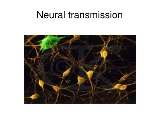
Ch 4: Neural Conduction & Synaptic Transmission. This chapter is about introducing the function of neurons How they conduct & transmit electrochemical signals through the nervous system. Resting Membrane Potential. Function of neurons centers around the membrane potential

E N D
This chapter is about introducing the function of neurons • How they conduct & transmit electrochemical signals through the nervous system
Resting Membrane Potential • Function of neurons centers around the membrane potential • The difference in electrical charge between the inside & outside of the cell • Can measure membrane potential using a microelectrode • Measure the charge inside the cell & the charge outside.
Resting Potential • A neuron’s resting potential is -70mV • Meaning, the charge inside the cell is 70mV less than the charge outside • Inside < Outside • Because this value is beyond 0, it is said to be polarized • So at rest, neurons are polarized.
Ionic Basis • It is polarized due to the arrangement of ions • The salts in neural tissues separate into + and – charged particles called ions • 4 main ions are responsible: • K+(potassium) • Na+(sodium) • Cl-(chloride) • - charged proteins
Ionic Basis • The ratio of – to + ions is greater inside a neuron than out, so you have a more – charge inside • Again, why the neuron’s resting potential is polarized • 2 things cause this imbalance & 2 things try to equalize (homogenize)
Contributing Factors to Resting Potential • Equalizers (homogenizers) • Random motion • Electrostatic pressure • Cause imbalance • Passive flow • Active transport
Equalizers • Random Motion • Ions are in constant random motion • Tend to be evenly distributed because they move down their concentration gradient • Move from areas of higher concentration to lower concentration • Electrostatic Pressure • Ions with the same charge will repel each other • Opposite charges attract
Contributing Factors to Resting Potential • Concentrations of Na+ and Cl- are greater outside the neuron (extracellularly) • K+ concentration is greater inside the cell (intracellularly) • Negatively charged proteins generally stay inside the neuron
Imbalance…rs • Passive Flow • Does not require energy • The membrane is selectively permeable to the different ions • K+ and Cl- ions easily pass through the membrane • Na+ ions have difficulty passing through • Ions passively flow across the membrane viaion channels • Special pores in the membrane • Active transport • Needs energy to power the pumps
Imbalance…rs • Active transport • Requires energy to power the pumps that transport the ions • Discovered by Hodgkin & Huxley • Nobel prize winning research • Why is there high Na+ and Cl- outside and high K+ inside? Why are they not passively flowing down their concentration gradients & reaching equilibrium? • Calculated the electrostatic pressure (mV) that would be necessary to counteract the passive flow down the concentration gradient (aka keep the concentrations uneven across the membrane) & how this differed from the actual resting potential
Active pumps cont. • Discovered that there are active pumps that counteract the passive flow of ions in & out of the cell (specifically for Na+ and K+) • Sodium-potassium pumps • Actively (using energy) pumps Na+ out & K+ in • 3 Na+ ions out for every 2 K+ ions pumped in • Other types of active transporters also exist *Summary Table 4.1 (pg. 79)*
Postsynaptic Potentials • Remember: At a synapse, the presynaptic neuron releases NT that bind with receptors on the postsynaptic neuron, to transmit the signal from one neuron to the next • When the NT bind with the postsynaptic neuron, they have either of 2 effects • Depolarize the membrane • Decrease the resting potential • **this means become less negative, aka approach zero** • Hyperpolarize the membrane • Increase the resting potential • ** make it more negative; further from zero**
0 mV depolarize -70 mV hyperpolarize
Postsynaptic Potentials • Postsynaptic depolarizations: • Excitatory postsynaptic potentials • EPSPs • Increase the likelihood that the neuron will fire • Postsynaptic hyperpolarizations: • Inhibitory postsynaptic potentials • IPSPs • Decrease the likelihood that the neuron will fire • Graded responses • Weak signals cause small PSPs; strong signals cause large PSPs
PSPs • Travel passively • Very rapid (practically instantaneous) • Like a cable • Deteriorate over distance • Lose amplitude as they go along • Fade out • Like sound
Integration of PSPs • Individual PSPs have almost no effect on getting a neuron to fire • However, neurons can have thousands of synapses on them & combining the PSPs from all of those can initiate firing • Called integration • Add all the EPSPs + IPSPs • Remember: • PSPs are graded & have different strengths • ExcitatoryPSPs increase the likelihood of firing & InhibitoryPSPs decrease the likelihood
Integration of PSPs • Neurons integrates PSPs in 2 ways • Over space: spatial summation • EPSP + EPSP = big EPSP • EPSP + IPSP = 0 (cancel each other out; assuming of equal strength) • IPSP + IPSP = big IPSP • Over time: temporal summation • 2 PSPs in rapid succession coming from the same synapse can produce a larger PSP
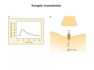
Action Potentials • If the sum of the PSPs reaching the axon hillock area at any one time is enough to reach the threshold of excitation, an action potential is generated • The threshold is -65mV • So the resting membrane potential must be depolarized 5mV for the neuron to fire • Action potential • Massive, 1ms reversal of the membrane potential • -70 to +50mV • Not graded; they are all-or-nothing responses • Either fire at full force or don’t fire at all
Conduction of APs • APs are generated & conducted via voltage-activated ion channels • When the threshold of excitation is hit, the voltage-activated Na+ channels open & Na+ rushes in • The Na+ influx causes the membrane potential to spike to +50mV • This triggers the voltage-gated K+ channels to open & K+ flows out • After 1ms, Na+ channels close • End of rising phase
Conduction of APs cont. • Beginning of repolarizing phase • K+ continues to flow out until the cell has been repolarized; then the K+ channels close • Cell returns to baseline resting membrane potential
Refractory Periods • For about 1-2ms after the AP, it is impossible to fire another one • Absolute refractory period • Followed by a period during which another AP can be fired, but it requires higher than normal levels of stimulation • Relative refractory period • Afterwards, the neuron returns to baseline & another AP can be fired as usual
Conduction in Myelinated Axons • Ions can pass through the membrane at the nodes of Ranvierbetween myelinsegments • APs move instantly through myelinated segments to the next node, where concentrated Na+ channels allow the signal to be “recharged” and sent to the next
Saltatory Conduction • Overall, this allows APs to be conducted much faster than in unmyelinated axons, because the AP “jumps” from node to node and effectively “skips” the lengths covered in myelin (saltatory conduction)
Velocity of Axonal Conduction • Speed of conduction is faster with myelin • Faster in thicker axons • Ex: mammalian motor neurons are thick & myelinated & can conduct signals at around 224 mph!!
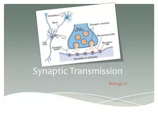
Structure of Synapses • Different types of synapses based on the location of the connection on each neuron • Axodendritic • “Normal” synapses • Terminal button of axon on Neuron1 to dendritic spine of Neuron2 • Axosomatic • Axon of N1 to soma of N2 • Dendrodendritic • Axoaxonic
Neurotransmitters • 2 categories of NTs • Large: • Neuropeptides • Small: • Made in terminal buttons & stored in vesicles
Release of NTs • NTs are released via exocytosis • At rest, NTs are in vesicles near membrane of presynaptic neurons • When an AP reaches the terminal button, voltage-activated Ca2+ channels open & Ca2+ rushes in • Ca2+ causes the vesicles to fuse with the membrane & release contents into the synaptic cleft
Activation of Receptors by NTs • NTs released from the presynaptic neuron cross the cleft & bind to receptors on the postsynaptic neuron • Receptors contain binding sites for only certain NTs • Any molecule that binds is a ligand • There are often multiple receptors that allow one kind of NT to bind: receptor subtypes • Different subtypes can cause different reactions
Receptors • There are 2 general types of receptors • Ionotropic • NT binds & ion channel opens & ions flow through • Immediate reaction • Metabotropic • NT binds & initiates a G-protein to trigger a second messenger, which moves within the cell to create a reaction • Slow, longer lasting effects • More abundant
Autoreceptor • A special type of metabotropic receptor • Located on the presynaptic neuron & bind with NTs from its own neuron • Function to monitor the # of NTs in the synapse • If too few, signal to release more • Too many, signal to slow/stop release
Reuptake, Degradation & Recycling • In order to allow the synapses to be available to signal again, the extra NT in the synaptic cleft need to be “cleaned up” by: • Reuptake • Most of the extra NT are quickly taken back into the presynaptic neuron by transporters to be repackaged in vesicles for future release • Enzymatic degradation • NTs in the cleft are broken down by enzymes • Ex: acetylcholine broken down by acetylcholinesterase • Even these pieces are taken back into the neuron & recycled
Gap Junctions • Unique signal transmission alternative to traditional synapses • Called electrical synapses • Narrow gaps between neurons connected by fine tubes called connexinsthat let electrical signals pass • Very fast & allow communication in both directions • Not yet fully understood in mammalian systems
Neurotransmitters • Amino Acid NTs • Monoamine NTs • Acetylecholine • Unconventional/Misc. NTs • Neuropeptides
Amino Acid NTs • AAs are the building blocks of proteins • Glutamate • Most common excitatory NT in the CNS • Aspartate • Glycine • GABA • Most common inhibitory NT
Monoamines • 2 groups with a total of 4 NTs in this class • Catecholamines: • Dopamine (DA) • Made from tyrosine/L-Dopa • Norepinephrine (NE) • Made from dopamine • Epinephrine • Made from NE • Indolamines: • Serotonin (5-HT) • Made from tryptophan
Acetylcholine (Ach) • Functions at neuromuscular junctions, in ANS & CNS • Extra is mostly broken down in the synapse; by acetylcholinesterase • Receptors for Ach are said to be cholinergic
Unconventional/Misc. NTs • Act differently than traditional NTs • Nitric oxide & carbon monoxide • Gases that diffuse across the membrane, across the extracellular fluid & across the membrane of the next neuron • Endocannabinoids • Essentially, the brain’s natural version of THC (main active chemical in marijuana) • Ex: annandimide
Neuropeptides • Don’t worry about the specific types • Just know that they are another type of NT • Generally large NTs
Drugs & Synaptic Transmission • Pharmaceutical drugs generally affect synaptic in 2 ways • Agonists facilitate the effects of a NT • Can bind to a receptor & activate it like the NT would • Antagonists inhibit • Can bind to a receptor & block it so NTs cannot bind
Example • Acetylcholine has 2 types of receptors • Nicotinic • Many in the PNS between motor neurons & muscle fibers • Ionotropic • Nicotine: agonist • Curare: antagonist (causes paralysis) • Botox: antagonist • Muscarinic • Many located in the ANS • Metabotropic • Atropine: antagonist, receptor blocker
Misc. • Endogenous • Compounds naturally made within the body • Ex: enkephalins & endorphins • The body’s endogenous opioids • An exogenous opioid is morphine • Opioids are analgesics (pain relievers)