Download
1 / 79
800 likes | 984 Vues
Cardiovascular Nursing Selected Topics PT 1. It’s All About Cardiac Output. Learning Outcomes a.k.a. “What’s On the Test?”. See the Study Guides! Setting the Stage / Context for Cardiac Monitoring & EKGs AAA “It’s All About Cardiac Output”—EKGs “It’s All About Cardiac Output”--Shock

E N D
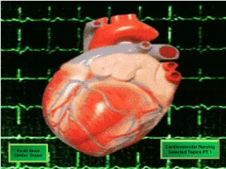
Cardiovascular Nursing Selected Topics PT 1 It’s All About Cardiac Output
Learning Outcomesa.k.a. “What’s On the Test?” • See the Study Guides! • SettingtheStage / Context for Cardiac Monitoring & EKGs • AAA • “It’s All About Cardiac Output”—EKGs • “It’s All About Cardiac Output”--Shock • Cardiac Monitoring: Selected Dysrhythmias, Recognition, Assessment, Nursing Interventions,
Selected Topics in Cardiovascular Nursing_____ ASSESSMENT Physical Assessment Inspection Palpation Percussion Auscultation Cardiac Monitoring Lab Monitoring PHARMACOLOGY Cardiac Glycosides ACE Inhibitors Beta Blockers Antiarrhythmics Catecholamines Anticoagulants PATHOPHYSIOLOGY Myocardial Infarction Acute Coronary Syndrome Valvular Heart Disease Pacemakers CABG Abdominal Aortic Aneurysm Pericarditis Peripheral Vasc Disease (PVD) Fem-Pop Bypass Graft Shock / Fluid Deficit Raynaud’s Phenomenon Arrhythmias / Dysrhythmias Care Planning Plan for client adl’s, Monitoring, med admin., Patient education, more… Nursing Interventions & Evaluation Execute the care plan, evaluate for Efficacy, revise as necessary
Remember: It’s All About Cardiac Output Cardiac Output = HR X SV C.O. = Heart Rate x Stroke Volume *More about this in Adult Health II
It’s All About… Cardiac Output What Factors Affect Cardiac Output ? Cardiac Output = HR X SV C.O. = Heart Rate x Stroke Volume *More about this in Adult Health II
It’s All About… Cardiac Output What Factors Affect Cardiac Output ? Sympathetic Nervous System (+) Cardiac Output = HR X SV C.O. = Heart Rate x Stroke Volume Parasympathetic Nervous System (-) *More about this in Adult Health II
It’s All About… Cardiac Output What Factors Affect Cardiac Output ? Sympathetic Nervous System Baroreceptors Cardiac Output = HR X SV Chemoreceptors C.O. = Heart Rate x Stroke Volume Parasympathetic Nervous System *More about this in Adult Health II
It’s All About… Cardiac Output What Factors Affect Cardiac Output ? Sympathetic Nervous System Baroreceptors Cardiac Output = HR X SV Blood Volume Chemoreceptors C.O. = Heart Rate x Stroke Volume Viscosity Of Blood Parasympathetic Nervous System Blood Volume *More about this in Adult Health II
It’s All About… Cardiac Output What Factors Affect Cardiac Output ? Medications Sympathetic Nervous System Medications Baroreceptors Cardiac Output = HR X SV Blood Volume Chemoreceptors C.O. = Heart Rate x Stroke Volume Viscosity Of Blood Condition of Heart Valves Parasympathetic Nervous System Blood Volume *More about this in Adult Health II
It’s All About… Cardiac Output What Factors Affect Cardiac Output ? Medications Sympathetic Nervous System Medications Baroreceptors Cardiac Output = HR X SV Blood Volume Chemoreceptors Preload, Afterload C.O. = Heart Rate x Stroke Volume Condition Of Myocardium Condition of Cardiac Conduction System* Viscosity Of Blood Condition of Heart Valves Parasympathetic Nervous System Blood Volume *More about this in Adult Health II
It’s All About… Cardiac Output What Factors Affect Cardiac Output ? Medications Sympathetic Nervous System Medications Baroreceptors Cardiac Output = HR X SV Blood Volume Chemoreceptors Preload, Afterload C.O. = Heart Rate x Stroke Volume Condition Of Myocardium And, Many more factors ! It’s All About CardiacOutput ! Condition of Cardiac Conduction System* Viscosity Of Blood Condition of Heart Valves Parasympathetic Nervous System Blood Volume *More about this in Adult Health II
Cardiovascular Nursing Selected Topics PT 1 Atrial Fibrillation Complicated Patient
Page R. 78 y.o. Sick Sinus Syndrome S/P Pacemaker Insertion Renee C. 29 y.o. Pericarditis Admission Pending Pre-Op CABG CARDIAC MONITORING Haynes H. 55 y.o. PVD S/P Femoral-Popliteal Bypass Kam H. 48 y.o. AAA James H. 68 y.o. R/O MI , Atrial Fibrillation
Treatments V.S. & Graphics Reports MISC I & O Assessments Consults Nurse’s Notes History & Physical Dr’s Orders Labs & Dx M.A.R. Patient Record Name: James H. Age: 68 y.o Male Occupation: Architect Adm: 11 Feb 2009 DX: R/O MI, R/O CVA, S/P CABG X 4 (1/22/2009) OTHER DX: DM, AAA, PVD, Atrial Fibrillation James H. Name:
Dr’s Orders • Admit to Telemetry Unit; continuous cardiac monitoring • DX: R/O MI, R/O Embolic CVA • Activity: BR, BSC • Diet: Clear Liquids, adv as tol to 1500 calorie ADA Diet • FSBG q ac & hs with Moderate SSRI Coverage • Meds: • Humulin 70/30 35 units sq q am / 20 units sq q pm • Digoxin 0.250 mg po daily • Amiodarone 400 mg po bid • Colace 200 mg po daily • Heparin IV per weight-based protocol • NTG 0.4 mg sl q5 min x 3, PRN CP • Morphine SO4 2 mg IVP PRN CP • Lidocaine 2 mg / minute IV / continuous • IV: Saline Lock • Labs / Diagnostics: Continue Serial Cardiac Enzymes; BMP q day; CBC; Coag studies per heparin weight-based protocol; schedule for CT of brain • Telemetry Protocols; ACLS Protocols • 10. Daily EKG; EKG with any chest pain What are your concerns about the patient? What is the cause of the concern? What are you going to do about it? What is the patient experiencing?
History & Physical SEE CONCEPT MAP Admitted 2/10/2009 after c/o crushing, substernal chest pain rated as 9 on a scale of 1-10.(Presented with Cardiac Rhythm as noted on ( ER rhythm strip #1 and ER rhythm strip #2 ); ( later, developed (Rhythm strip #3) while being transported from the Emergency Department to the telemetry unit.) Also was noted to have rhythm noted on Rhythm strip #4.Rhythm strip #5 is attached for your enlightenment. Client was successfully resuscitated, including use of ACLS protocols and Defibrillation with 360 joules x 2. Converted to atrial fibrillation w/controlled ven-tricular response. After defibrillation and transfer to the nursing unit, pt exhibited s/s disorientation-see Rhythm Strip #6. Five hours after admission to the telemetry floor, became agitated and c/o (R)-side chest pain: See Nurse’s Notes. Surgical history includes 4 vessel CABG in 1/2008; PTCA with 3 stents in 2002; LaparascopicCholecystectomy in 1999. Other pertinent Medical History includes diagnosis of DM in 1990; blood sugars controlled moderately well with Humulin 70/30 35 units q am / 20 units q pm. Long history of atrial fibrillation with concomitant control via digoxin 0.25 mg daily. CO = HR & R X SV BP = CO X SVR
RHYTHM STRIP #1 • The Patient airway, breathing, LOC Awake and alert; BP= 112/72
RHYTHM STRIP #2 Awake & Alert C/O Chest Pressure and Feeling Nervous BP = 106/68 • Check The Patient airway, breathing, LOC
RHYTHM STRIP #3 The Patientairway, breathing, LOC Awake and alert; BP= 88/40 C/O “Feeling Funny”
RHYTHM STRIP #4 The Patient (!)airway, breathing, LOC NON-RESPONSIVE BP= CO = HR&R x SV BP = CO x SVR
RHYTHM STRIP #5 AWAKE & ALERT BP = 112/78 CHEST LEAD RECONNECTED (It fell off…) (Oops, MY BAD!) • The Patient (!) Airway, breathing, LOC TREAT THE PATIENT, NOT THE MONITOR !
RHYTHM STRIP # 6 The Patientairway, breathing, LOC BP = 112/72 Speech slurred (Check cranial nerves) II Optic III Oculomotor IV Trochlear V Trigeminal VI Abducens VII Facial IX, X Glossopharyngeal Vagus
What are your concerns about the patient? What is the cause of the concern? What are you going to do about it? What is the patient experiencing? Nurse’s Notes SEE CONCEPT MAP 0700: Unable to initiate additional peripheral IV line, attempts x 3. VS: P=90, irregular; R=22, unlabored; BP= 118/78; SaO2=95%. Monitor displaying atrial fibrillation with occasional PVC. Physician notified re: IV; will continue Lidocaine gtt @ 2 mg/min per available site on (L) forearm; Heparin infusion per WBP on hold until additional IV site accessed. Consult for central line placement pending.------------------------------------------------------------------------------J. Nurse, RN 0900: C/O sharp pain, pointing to area (R) thorax; became agitated and dis- oriented . VS: BP = 90/50; P = 110, irregular; R = 32, labored; T =98*; SaO2 = 86 %. ABG’s obtained, results pending. O2 increased to 4L/NC. Note absent breath sounds, RLL;Cardiac monitor: Atrial fibrillationw/ rapid ventricular response. Physician notified and enroute to hospital. Will continue to Monitor.---------------------------------------------------------------------------J. Nurse, RN 0920: Non-responsive; VS: BP = 80/40, P = 156, irreg, R = 10, SaO2 = 80%; central cyanosis noted. Intubated and ventilated with 100% 02. Absent lung sounds RLL & RML. Cardiac monitor shows atrial fibrillationw/ uncontrolled ventricular response. Report provided to ICU nurse, Transported via gurney to ICU for ventilator support.-----------------------J. Nurse, RN
NSG DX #3: Ineffective Tissue Perfusion, Cerebral NSG DX #2: Ineffective Tissue Perfusion, Cardio- pulmonary NSG DX #4: Impaired Gas Exchange NSG DX #1: PAIN, ACUTE MED DX:MI Atrial Fib CVA PE Other Nursing DX: 4. Decreased Cardiac Output 5. IMPAIRED SWALLOWING 6. RISK for INJURY 7. Self-Care Deficit 8. Impaired Communication KEY ASSESSMENTS PAIN VS O2 Sats ABG’s LOC Cranial Nerve Assmt Capillary Refill Breath Sounds Swallowing / Gag Reflex CONCEPT MAP James H.
Cardiovascular Nursing Selected Topics PT 2 Abdominal Aortic Aneurysm
AAA Patient Record NAME: Kam H. AGE: 48 y.o. OCCUPATION: Attorney ADM: 2/11/2008 DX: AAA Pre-Op: AAA Repair 2/12/2008 NAME: Kam H.
S/S FREQUENTLY: ASYMPTOMATIC Gnawing, constant, abdominal, flank, and groin pain Pulsating abdominal mass Bruit RUPTURE Sudden onset “tearing,” “ripping,” or “stabbing” abdominal or back pain Shock (Hypovolemic) GRAFT OCCLUSION Changes in Pulses Coolness & cyanosis of extremities below graft Severe Pain Decreased Urine Output AAAAbdominal Aortic Aneurysm
Nursing Care:AAA Repair ? ? NSG DX #1: Fear / Anxiety ? Abdominal Aortic Aneurysm (Pathophysiology) Other Nursing Diagnoses That May Apply: ? KEY ASSESSMENTS?
AAA: Pathophysiology • An abdominal aortic aneurysm is an abnormal dilation of the wall of the abdominal aorta. The aneurysm usually develops in the segment of the vessel that is between the renal arteries and the iliac branches of the aorta. The most common cause of an abdominal aortic aneurysm is atherosclerosis. The plaque that forms on the wall of the artery causes degenerative changes in the medial layer of the vessel. These changes lead to loss of elasticity, weakening, and eventual dilation of the affected segment. Some other causes of abdominal aortic aneurysm include inflammation (arteritis), trauma, infection, congenital abnormalities of the vessel, and connective tissue disorders that cause vessel wall weakness. • Most abdominal aortic aneurysms are asymptomatic and are discovered during a routine physical examination (signs include palpation of a pulsatile mass in the abdomen and/or auscultation of a bruit over the abdominal aorta) or during a review of x-ray results of the abdomen or lower spine. The presence of symptoms such as mild to severe abdominal, lumbar, or flank pain and/or lower extremity arterial insufficiency is usually indicative of a large aneurysm that is exerting pressure on surrounding tissues or an aneurysm that is leaking. • Surgical repair of an aneurysm is usually performed if the aneurysm is growing rapidly and/or reaches a size of 5-6 cm or larger or if the client experiences symptoms. The procedure often involves the use of a synthetic graft, which is inserted to replace or support the weakened vessel. Ulrich & Canale: (2006) Nursing Care Planning Guides: For Adults in Acute, Extended, and Home Care Settings, 6th Edition
NURSING DIAGNOSIS: Fear/Anxietyrelated to: • 1. unfamiliar environment and separation from significant others; • 2. lack of understanding of diagnostic tests, surgical procedure, and postoperative care; • 3. anticipated loss of control associated with effects of anesthesia; • 4. risk of disease if blood transfusions are necessary;5. anticipated postoperative discomfort and potential change in sexual functioning; • 6. possibility of death. • Desired OutcomeThe client will experience a reduction in fear and anxiety • Nursing Actions and Selected Purposes/Rationales1. Preoperative Care Plan, for measures related to the assessment and reduction of fear and anxiety.2. Implement additional measures to reduce fear and anxiety: a. orient client to critical care unit if appropriate b. describe and explain the rationale for equipment and tubes that may be present postoperatively (e.g., cardiac monitor, ventilator, intravenous and intra-arterial lines, nasogastric tube, urinary catheter) c. explain that B/P may be taken in both arms and thighs in order to better evaluate circulatory status d. reinforce physician's explanations and clarify misconceptions client has about effects of the surgery on sexual functioning (impotence can result from diminished blood flow in the mesenteric or internal iliac arteries during or after surgery and/or from nerve damage during surgery).
COLLABORATIVE DIAGNOSIS: Potential complication: hypovolemic shockR/T related to excessive blood loss if the aneurysm ruptures. • Desired Outcome:The client will not develop hypovolemic shock as evidenced by:1. usual mental status2. stable vital signs3. skin warm and usual color4. palpable peripheral pulses5. urine output at least 30 ml/hour. • NURSING ACTIONS: • (next page)
Nursing Actions and Selected Purposes/Rationales • 1. Assess for and immediately report signs and symptoms of conditions that indicate impending aneurysm rupture: • A. Leaking aneurysm: a. increasing abdominal girth b. ecchymosis of flank area or perineum c. frank or occult gastrointestinal bleeding (occurs if the aneurysm ruptures into the duodenum) d. decreasing RBC, Hct, and Hgb levels e. new or increased reports of lumbar, flank, abdominal, pelvic, or groin pain (accumulation of blood in the peritoneum and/or retroperitoneal spaces causes irritation of and pressure on the tissues and nerves) f. diminishing or absent peripheral pulses g. further decline in thigh B/P as compared with B/P in arm (thigh B/P is usually slightly lower than B/P in arm of a client with an abdominal aortic aneurysm) • B..Expanding aneurysm: a. new or increased reports of lumbar, flank, or groin pain (results from pressure on lumbar nerves) b. increased size of pulsating mass in abdomen c. increasing sense of abdominal and/or gastric fullness (results from pressure on duodenum) d. decreasing motor or sensory function of lower extremities (results from pressure on lumbar and/or sacral nerves). • C. Assess for and report signs and symptoms of hypovolemic shock: a. restlessness, agitation, confusion, or other change in mental status b. significant decrease in B/P c. postural hypotension d. rapid, weak pulse e. rapid respirations f. cool skin g. pallor, cyanosis h. diminished or absent peripheral pulses i. urine output less than 30 ml/hour.
D. Implement measures to decrease risk of aneurysm rupture: a. instruct client to avoid elevating legs when in bed, using knee gatch, and crossing legs in order to prevent restriction of blood flow to the lower extremities and subsequent increase in vascular pressure at the aneurysm site b. perform actions to prevent an increase in blood pressure: c. limit client's activity as ordered d. nstruct client to avoid activities that create a Valsalva response (e.g., straining to have a bowel movement, holding breath while moving up in bed, lifting heavy objects) • E. implement measures to reduce fear and anxiety (see Preoperative Diagnosis 1) • F. administer antihypertensives if ordered to reduce pressure in the dilated vessel. • G. If signs and symptoms of hypovolemic shock occur: • a. place client flat in bed unless contraindicated b. monitor vital signs frequently c. administer oxygen as ordered d. administer blood and/or volume expanders as ordered (these need to be used with caution since increased vascular pressure can extend a tear at site of rupture) e. prepare client for insertion of hemodynamic monitoring devices (e.g., central venous catheter, intra-arterial catheter) if indicated f. prepare client for emergency surgical repair of aneurysm if indicated.
NURSING DIAGNOSIS: Risk for imbalanced fluid and electrolytes • Third-spacing of fluid related to: • 1. increased capillary permeability in surgical area associated with the inflammation that occurs following extensive dissection of tissue during major abdominal surgery • 2. increased vascular hydrostatic pressure associated with excess fluid volume if present • 3. hypoalbuminemia associated with the escape of proteins from the vascular space into the peritoneum (a result of increased capillary permeability in the surgical area); • Excess fluid volume related to: • 1. vigorous fluid replacement • Fluid retention associated with: • 1. increased secretion of antidiuretic hormone (output of ADH is stimulated by trauma, pain, and anesthetic agents) • 2. renal insufficiency (can occur if there is inadequate blood flow to the kidneys during or after surgery) • 3. reabsorption of third-space fluid (occurs about the 3rd postoperative day); • Deficient fluid volume related to restricted oral fluid intake before, during, and after surgery; blood loss; and loss of fluid associated with nasogastric tube drainage; • Electrolyte Imbalance: hypokalemia, hypochloremia, and metabolic alkalosis related to loss of electrolytes and hydrochloric acid associated with nasogastric tube drainage.
Desired OutcomeThe client will experience resolution of third-spacing as evidenced by:1. absence of ascites2. B/P and pulse within normal range for client and stable with position change.
Cardiovascular Nursing Selected Topics PT 3 Peripheral Vascular Disease
PVD Patient Record NAME: Haynes H. AGE: 55 y.o. OCCUPATION: Registered Nurse ADM: 2/9/2008 DX: Peripheral Vascular Disease Procedure: Femoral-Popliteal Bypass 2/10/2008 NAME: Haynes H.
Peripheral Arterial Disease • Pathophysiology • PAD results from atheroclerosis in the arteries of the lower extremities, characterized by inadequate blood flow (ischemia). • Intermittent Claudication: pain caused by insufficient arterial blood supply
PTA : Percutaneous Transluminal Angioplasty
An intraoperative photograph of a right femoral to posterior tibial artery bypass using the greater saphenous vein to correct peripheral arterial disease.
Nursing ConsiderationsPost Femoral-Popliteal Bypass MEDS • Hemorheologic Drugs: pentoxifyline (Trental) increases RBC flexibility, decreases viscosity • Antiplatelet Agents: ASA, clopidogrel (Plavix) • Pedal Pulses (palpated or Doppler) • Color, temp., capillary refill, pain (warmth, redness, & edema are EXPECTED OUTCOMES of the revascularlization). • Graft Occlusion NOTIFY PHYSICIAN • Compartment Syndrome NOTIFY PHYSICIAN • Acute Arterial Occlusion NOTIFY PHYSICIAN ASSESSMENT COMPLICATIONS THE 6 P’s of Ischemia: PAIN, PALLOR, PULSELESSNESS, PARESTHESIA, PARALYSIS, POIKILOTHERMIA
Buerger’s Disease thromboangiitis obliterans Raynaud’s Disease Raynaud’s phenomenon PAD
Pacemaker Patient Record NAME: Page R. AGE: 78 y.o. OCCUPATION: Retired Teacher ADM: 2/9/2008 DX: Sick Sinus Syndrome Procedure: Pacemaker Insertion 2/10/2008 NAME: Page R.
Pathophysiology Nursing Considerations Cardiac Pacemakers
IMMEDIATELY POST-OP: Monitor heart rate & rhythm Minimize shoulder movement w/ sling for 24 hrs; Gentle passive ROM after 24 hrs Indications: Symptomatic bradycardia Complete Heart Block Sick Sinus Syndrome Sinus arrest Asystole Atrial tachydysrhythmias Ventricular tachydysrhythmias Cardiac Pacemakers
Pacemaker Complications • Failure to Capture— Pacemaker initiates a stimulus, but depolarization of the myocardium does not occur Stimulation of Chest Wall or Diaphragm Hiccoughs Cardiac Tamponade