Algorithmic Competition for Liver Region Extraction from 3D CT Images in Japan
10 likes | 88 Vues
Preliminary report on liver region extraction algorithms competition - crucial for early detection of liver cancer. Detailed evaluation by radiologists using different methods. Future plans for assessing liver cancer detection performance.

Algorithmic Competition for Liver Region Extraction from 3D CT Images in Japan
E N D
Presentation Transcript
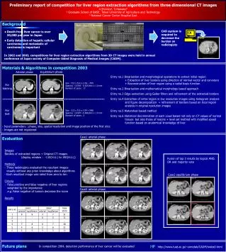
Tumor No.1 No.1 No.2 No.2 No.3 No.3 No.4 No.4 No.5 No.5 No.6 No.6 No.1 No.4 No.6 Fusion Preliminary report of competition for liver region extraction algorithms from three dimensional CT images A.Shimizua, S.Nawanob a Graduate School of BASE, Tokyo University of Agriculture and Technology b National Cancer Center Hospital East Background Multi slice CT Scanner CAD system is required to decrease the burden on radiologists CAD • Death from liver cancer is over 30,000 per year in Japan • Early detection of hepatic cellular carcinoma and metastatic of carcinoma is important In 2002 and 2003, competitions for liver region extraction algorithms from 3D CT images were held in annual conference of Japan society of Computer Aided Diagnosis of Medical Images (CADM). Materials & Algorithms in competition 2003 Equilibrium phase Arterial phase Entry no.1 Binarization and morphological operations to extract initial region + Extraction of liver borders using direction of normal vector and curvature + Reconstruction of liver region using a radial basis function Entry no.2 Binarization and mathematical morphology based approach Entry no.3 Edge extraction using Gabor filters and refinement of the extracted borders Entry no.4 Extraction of initial region in low resolution images using histogram analysis and figure decomposition + refinement of borders based on local region analysis in original resolution images Entry no.5 Watershed based method Entry no.6 Statistical discrimination at each voxel based not only on CT values of normal tissues but also those of lesions + level set method with modified speed function based on anatomical knowledge of liver Size : 512 x 512 x (154 - 267) Spacing : (0.546- 0.625)mm x 1.0 mm Number of cases : 17 For training For test Size : 512 x 512 x (197~248) Spacing : (0.587~0.606)mm x 1.0 mm Number of cases : 2 • Input parameters : phase, size, spatial resolution and image position of the first slice • Images are not registered Case1 arterial phase Evaluation • Images • Borders of extracted regions +Original CT images • (display window : -110(H.U.) to 190(H.U.)) • Methods • - Three radiologists evaluated the resultant images visually without any priori knowledge about algorithms • Each resultant image was rated from zero to ten • Criteria • False positive and false negative of liver regions • weighted by the importance • e.g. False negative of tumors decrease the score • Results Fusion of top 3 results by logical AND, OR and majority vote Case2 equilibrium phase Case2 arterial phase AND OR Majority vote Future plans HP http://www.tuat.ac.jp/~simizlab/CADM/index0.html In competition 2004, detection performance of liver cancer will be evaluated


