Download
1 / 41
422 likes | 669 Vues
Genes of cancer. Cancer is a disease of abnormal cells Cancer cells proliferate in an uncontrolled fashion The causes of cancer are quite diverse but the central role is played by DNA mutations in development of cancer. Chemicals. spontaneous. Oncogenes. radiation. Mutation. Cancer.

E N D
Genes of cancer • Cancer is a disease of abnormal cells • Cancer cells proliferate in an uncontrolled fashion • The causes of cancer are quite diverse but the central role is played by DNA mutations in development of cancer
Chemicals spontaneous Oncogenes radiation Mutation Cancer Tumour suppressor genes infection Errors in DNA replication
Cancer related mutations affect the same two classes of genes : Oncogenes and tumour suppressor genes • Oncogenes are defined as genes whose presence can lead to cancer. They arise by mutation from normal genes (proto oncogenes) that code for proteins involved in stimulating cell proliferation and survival. By producing abnormal forms or excessive quantities of these proteins, oncogenes contribute to the uncontrolled proliferation and survival of cancer cells. • In contrast to oncogenes which are abnormal genes, tumour suppressor genes are normal genes whose deletion or loss function can likewise lead to cancer. Tumour suppressor gene produce proteins that either directly or indirectly exert a restraining influence on cell proliferation and survival. The loss of such proteins can therefore allow cell proliferation and survival to evade normal restraints and controls.
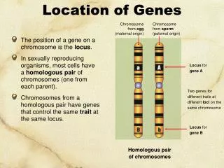
Genetic flow of information • Chromosomes within our cells have roughly 30,000genes. • Each gene codes for a RNA molecule that is either directly used or used as a guide for the formation of protein • DNA (store) to RNA (working form ) to protein (product)
THE FLOW OF GENETIC INFORMATION DNA RNA PROTEIN 1 2 3 DNA 1. REPLICATION (DNA SYNTHESIS) 2. TRANSCRIPTION (RNA SYNTHESIS) 3. TRANSLATION (PROTEIN SYNTHESIS)
RNA polynucleotide chain • 2’ -OH makes • 3’, 5’ phosphodiester • bond unstable DNA polynucleotide chain
Transcription • The process in which a particular section of DNA (genes) are used to produce RNA is known as transcription • Goal of transcription is to make an RNA copy of a gene. • Only a small percentage of genes are actually being used to make RNA at a particular time in a particular cell
The transcription process is tightly regulated in normal cells. • Genes must be transcribed at the correct time. • The RNA produced from the gene must be made in correct amount • Only the required gene must be transcribed. • Turning transcription off is just as important as turning it on.
The steps of transcription • A transcription factor recognises the start site (promoter) of a gene to be transcribed • The enzyme that makes RNA (RNA polymerase) binds to transcription factor and recognises the start region • The enzyme proceeds down the DNA making a copy until the end is reached. • The enzyme falls of and RNA is released. This copying process may be repeated numerous times • If the RNA is one that codes for protein it will leave the nucleus to enter the cytosol
transcription unit Transcription and promoter elements for RNA polymerase II +1 transcription element TE P exon exon promoter • Promoter (DNA sequence upstream of a gene) • determines start site (+1) for transcription initiation • located immediately upstream of the start site • allows basal (low level) transcription • Transcription element (DNA sequence that regulates the gene) • determines frequency or efficiency of transcription • located upstream, downstream, or within genes • can be very close to or thousands of base pairs from a gene • includes • enhancers (increase transcription rate) • silencers (decrease transcription rate) • response elements (target sequences for signaling molecules) • genes can have numerous transcription elements
Proteins regulating eukaryotic mRNA synthesis • General transcription factors • TFIID (a multisubunit protein) binds to the TATA box • to begin the assembly of the transcription apparatus • the TATA binding protein (TBP) directly binds the TATA box • TBP associated factors (TAFs) bind to TBP • TFIIA, TFIIB, TFIIE, TFIIF, TFIIH1, TFIIJ assemble with TFIID • RNA polymerase II binds the promoter region via the TFII’s • Transcription factors binding to other promoter elements and • transcription elements interact with proteins at the promoter • and further stabilize (or inhibit) formation of a functional • preinitiation complex • 1TFIIH is also involved in phosphorylation of RNA polymerase II, DNA repair • (Cockayne syndrome mutations), and cell cycle regulation
Initiation of transcription and promoter clearance F E B TFIID H initiation TBP J +1 RNA pol II CTD P P P • RNA pol II is phosphorylated by TFIIH on the carboxy terminal • domain (CTD), releasing it from the preinitiation complex and • allowing it to initiate RNA synthesis and move down the gene
Transcription (elongation) /antisense strand
Transcription (termination) RNA polymerase falls off terminator Coding strand 5’TACGCTGCCCAAGCA Template strand 3’ATGCGACGGGTTCGT RNA sequence Animation: http://www.phschool.com/science/biology_place/biocoach/images/transcription/tcani.gif
Transcription factors • The inappropriate activity of transcription factors has been identified in almost all types of known cancers, Some examples of transcription factors that malfunction in human cancers. • P53- The protein that the p53 codes foris important because it controls the transcription of genes involved in causing cells to divide. • Rb- The protein product of this gene works by blocking other transcription factors thus preventing transcription of key genes required for cell division to progress. • The oestrogen receptor (ER) This protein binds oestrogen and the combination acts as transcription factor to turn on genes that enable target cells to divide.
What is translation • After hnRNA production through the process of transcription, it is processed in the nucleus to produce mRNA which is then released into cytosol. • The mRNA is then recognised by the ribosomal subunits and the message is read by the ribosome to produce a protein. The information for the direction of protein synthesis is encoded in nucleotide sequence that makes up mRNA. Groups of three nucleotides (codons) are read by ribosomes and lead to the insertion of a particular amino acid in growing peptide. • After the protein is formed it is folded to perform its function in the cell The proper folding, transportation, activity and eventual destruction of protein are all highly regulated processes. The genes that control these processes are often found to be damaged and malfunction in cancer cells.
Messenger RNA (mRNA) initiation codon Cap 5’ untranslated region 5’ AUG m7Gppp translated (coding) region UGA termination codon 3’ untranslated region AAUAAA (AAAA)n 3’ poly(A) tail
Protein translation: summary Elongation Initiation Termination http://www.phschool.com/science/biology_place/biocoach/translation/init.html
Reading frame • reading frame is determined by the AUG initiation codon • every subsequent triplet is read as a codon until reaching a stop codon • ...AGAGCGGA.AUG.GCA.GAG.UGG.CUA.AGC.AUG.UCG.UGA.UCGAAUAAA... • MET.ALA.GLU.TRP.LEU.SER.MET.SER • a frameshift mutation • ...AGAGCGGA.AUG.GCA.GA .UGG.CUA.AGC.AUG.UCG.UGA.UCGAAUAAA... • the new reading frame results in the wrong amino acid sequence and • the formation of a truncated protein • ...AGAGCGGA.AUG.GCA.GAU.GGC.UAA.GCAUGUCGUGAUCGAAUAAA... • MET.ALA.ASP.GLY
Cell division and mitosis • For mitosis to take place the following must occur; • The genetic material , the DNA in chromosomes , must be faithfully copied. This occurs via a process known as replication • The organelle, such as mitochondria , must be distributed so that each daughter cell receives adequate amount to function • The cytoplasm of the cell must be physically separated into two different cells. • Many features of cancer cells are due to defects in the genes that control cell division.
Themammalian cell cycle DNA synthesis and histone synthesis Rapid growth and preparation for DNA synthesis S phase G0 G1 phase Quiescent cells G2 phase Growth and preparation for cell division M phase Mitosis
Overview of the major events in mitosis Interphase prophase metaphase anaphase telophase In case of DNA damage or failure of critical processes P53 stimulates induction of inhibitory proteins that halt DNA replication Defects in p53 are associated with a variety of cancers DNA damage repair or initiation of programmed cell death (apoptosis)
DNA synthesis • Occurs in the S-phase • Every chromosome is copied with high fidelity. • In this process double stranded DNA is unwound and each individual strand is used as a template for the production of complimentary strand. • Errors may occur during replication that lead to changes in the nucleotide sequence of the chromosomes. If these changes occur within genes they can alter function of the cell. Human cells have evolved several mechanism to correct errors of this type but they are not perfect. • These mistakes can lead to mutated genes. • Accumulation of mutations can lead to the development of cancer • There are several cancers types that are associated specifically with breakdown of repair processes.
DNA replication is semi-conservative Parental DNA strands Each of the parental strands serves as a template for a daughter strand Daughter DNA strands Daughter strand Parent strand 1 5’GATCCTAGGTACTGACCTTGC3’ Parent strand 2 3’CTAGGATCCATGACTGGAACG5’ Daughter strand
Features of DNA Replication • DNA replication is semiconservative • Each strand of template DNA is being copied. • DNA replication is bidirectional • Bidirectional replication involves two replication forks, which move in opposite directions • DNA replication is semidiscontinuous • The leading strand copies continuously • The lagging strand copies in segments (Okazaki fragments) which must be joined
Mechanisms of Repair • Mutations that occur during DNA replication are repaired when possible by proofreading by the DNA polymerases • Mutations that are not repaired by proofreading are repaired • by mismatched (post-replication) repair followed by • excision repair • Mutations that occur spontaneously and in response to mutagens at any time are repaired by excision repaired (base excision or nucleotide excision)
CH3 CH3 CH3 CH3 Mismatched (post-replication) repair • the parental DNA strands are • methylated on certain • adenine bases • mutations on the newly • replicated strand are • identified by scanning • for mismatches prior to • methylation of the newly • replicated DNA 5’ 3’ • the mutations are repaired • by excision repair mechanisms • after repair, the newly • replicated strand is methylated
Some common type of DNA damage • Depurination involves loss of the base adenine or guanine caused by hydrolysis of the bond linking it to DNA chain • Deamination involves the removal of an amino group by hydrolysis • Pyrmidine dimers are created by an environmental mutagen, the UV radiation in sunlight
Deamination of cytosine can be repaired Deamination of 5-methylcytosine cannot be repaired More than 30% of all single base changes that have been detected as a cause of genetic disease have occurred at 5’-mCG-3’ sites
Excision repair (base or nucleotide) deamination ATGCUGCATTGA TACGGCGTAACT uracil DNA glycosylase ATGCGCATTGA TACGGCGTAACT repair nucleases AT GCATTGA TACGGCGTAACT DNA polymerase b ATGCCGCATTGA TACGGCGTAACT DNA ligase ATGCCGCATTGA TACGGCGTAACT thymine dimer ATGCUGCATTGATAG TACGGCGTAACTATC excinuclease AT AG TACGGCGTAACTATC (~30 nucleotides) DNA polymerase b ATGCCGCATTGATAG TACGGCGTAACTATC DNA ligase ATGCCGCATTGATAG TACGGCGTAACTATC Base excision repair Nucleotide excision repair
Defects in DNA repair or replication • Xeroderma pigmentosum • Ataxia telangiectasia • Fanconi anemia • Bloom syndrome • Cockayne syndrome 100 human elephant cow Life span 10 hamster Correlation between DNA repair activity in fibroblast cells from various mammalian species and the life span of the organism rat mouse shrew 1 DNA repair activity
The control of cell division. • Is the DNA fully replicated? • Is the DNA damaged? • Are there enough nutrients to support cell growth • If these checks fail normal cells will stop dividing • Cancer cells do not obey these rules and will continue to grow and divide.
The control of cell division • Cells divide in response to external signals • What are the signals that make cells stop dividing • A lack of positive external signals • Contact inhibition • Cellular Senescence
Cell division in cancer cells • Cancer cells can divide without appropriate external signals • Cancer cells do not exhibit contact inhibition • Cancer cells divide without receiving the ‘all clear’ signal.