Download
1 / 11
120 likes | 176 Vues
Explore potential causes of airway obstruction post-anesthesia induction and learn about devices to restore airway patency. Discover predictors of difficult mask ventilation and how to approach anticipated difficult intubation scenarios.

E N D
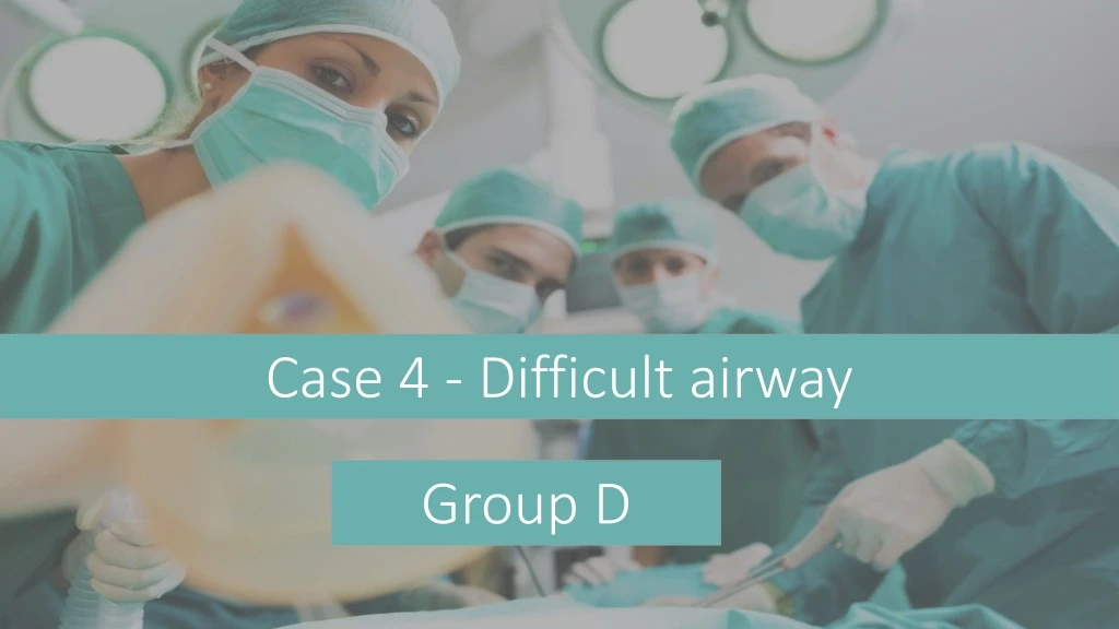
Case 4 - Difficult airway Group D
A 35-year-old woman presented for laparoscopic lysis of adhesions. Her first laparotomy occurred 10 years prior to this admission. After induction of anesthesia the patient had airway obstruction.At that time, the process of tracheal intubation consumed 1 hour. She awakened with a very sore throat, but she does not know the details of the intubation.
What is the causes of airway obstruction after induction of general anesthesia? • Atelectasis:Major cause of post operative hypoxia is atelectasis. • Ventilation associated:pneumonia is associated with the placement of an endotracheal tube and mechanically assisted ventilation. Noninvasive mechanical ventilation may help reduce the risk of pneumonia. • Aspiration Pneumonitis:Regurgitation and aspiration of gastric contents during induction of anaesthesia leads to aspiration pneumonitis. • CNS respiratory depression:affect the central regulation of breathing, changing the neural drive to respiratory muscles such as the diaphragm.
local Inflammatory Response: reflex stimulation during airway instrumentation and release of inflammatory mediator. • Increased salivary and respiratory secretions: Anesthetic gasses and tracheal intubation may impair normal muco- ciliary transport. • Negative Pressure Pulmonary Edema. • Pneumothorax. • Pulmonary Embolus. • Disrupt normal coordination of respiratory muscle action. • Postoperative pain: may cause voluntary limitation of respiratory motion. • Laryngeal and pharyngeal muscle relaxation.
What are the devices can be used to restore airway patency? • In most cases, only simple methods of airway clearance are required (e.g. use a head-tilt and chin-lift maneuver to open the airway, airways suction for blood or secretions, insertion of an oropharyngeal or nasopharyngeal airway( • Provide high-concentration oxygen using a mask with oxygen reservoir. • Endotracheal Intubation in emergency situations: • Naso-tracheal intubation • Oro-tracheal intubation • Surgical routes: (Cricothyroidotomy and Emergent Tracheostomy) REMEMBER: DO NOT insert your finger into a patients mouth and take good care of a patient who has lose or crowned teeth.
What are the predictors of difficult mask ventilation? * previous history* BOOTS: * Patient with active airway obstruction (tumor, abscess, laryngeal edema)
How is the anticipated difficult intubation approached? • Tracheal intubation non-essential ➡️supraglottic device ➡️intubation with the patient awake • In certain cases, a sevoflurane induction may be chosen to test the efficacy of a supraglottic device. • Tracheal intubation required ➡️supraglottic device may be used as a bridge.
Describe the management options for a patient who can’t ventilate cant intubate ? • Surgical airway (crico-thyroid puncture / tracheostomy). • tracheostomy can performed electively under local anesthesia for some surgeries such as large upper airway cancers.
How you can verify successful tracheal intubation? • Physical examination methods such as auscultation of chest and epigastrium, visualization of thoracic movement, and fogging in the tube ARE NOT sufficiently reliable to confirm endotracheal tube placement. • pulse oximetry and chest radiography ARE NOT reliable as sole techniques to determine endotracheal tube location. • So, you confirm it by : • direct visualizationof the endotracheal tube passing through the vocal cords into the trachea( especially with the use of a videolaryngoscope). • Use an end-tidal carbon dioxide detector (continuous waveform capnography, colorimetric and non-waveform capnography) in patients who have adequate tissue perfusion. • Use esophageal detector device, ultrasound, or bronchoscopy for patients in cardiac arrest and for those with markedly decreased perfusion.
References: • POSTOPERATIVE PULMONARY COMPLICATIONS Dr. Rudra A. Dr. SudiptaDas. • Preventing Postoperative Pulmonary Complications. David O. Warner, • Management of the anticipated difficult airway—a systematic approach: Continuing Professional Development, Pierre Drolet • Anaesthesiaat a Glance. Julian Stone, William Fawcett.
THANK YOU • Anjod Mansour Almuhareb • RahmaAbdulrahmanAlshehri • Sara Adnan Habis • HalaEbrahim Al-Askar • NuhaHamadAlhomayed • Albatoul Abdullah Alsuhaibani • Razan Abdullah Aldhahri • KholoudHushaish Al-Baqmi • Amjad Ali Abalkhail • AreejessaAlwehaib • Sarah Nasser Alshehri • Fatimah DhaferAlQarni