Download
1 / 330
3.3k likes | 3.54k Vues
Interrelationships Between Cerebral Infarction And Carotid Atherosclerosis With Some Risk Factors A thesis submitted to the College of Medicine, University of Al-Mustansiriya in partial fulfilment for the requirements of the degree of Doctor in Philosophy in Physiology

E N D
Interrelationships Between Cerebral Infarction And Carotid Atherosclerosis With Some Risk Factors A thesis submitted to the College of Medicine, University of Al-Mustansiriyain partial fulfilment for the requirements of the degree of Doctor in Philosophy in Physiology ByBasim M.H. ZwainB.D.S. (Bag), M.Sc. (Kufa)Supervised byProfessor Dr. and Professor Dr. Bassam T.MF. Al-Gailani Yesar MH. Al-Shamma’a M.B.Ch.B.(Bag) M.B.Ch.B.(Bag)Ph.D.(Leeds, UK) Ph.D.(Leeds, UK) START
Supervisor Professor Doctor Bassam T.MF. Al-Gailani M.B.Ch.B. (Baghdad), Ph.D. (Leeds, UK). Head Department of Physiology College of Medicine University of Al-Mustansiriya EXIT
Supervisor Professor Doctor Yesar M.H. Al-Shamma’a M.B.Ch.B. (Baghdad), Ph.D. (Leeds, UK).Head Department of Physiology College of Medicine University of Kufa EXIT
Student Basim M.H. Zwain B.D.S. (Baghdad) M.Sc. Physiology (Kufa) EXIT
WELCOME DO YOU WANTTO: VIEW ITEMS ? ? ? VIEW PAGES EXIT OR
ABSTRACT INTRODUCTION SUBJECTS AND METHODS RESULTS DISCUSSION REFERENCES MISCELLANEOUS EXIT HOME
HELLO AGAIN NOW, DO YOU WANT TO: VIEW ITEMS ? ? ? VIEW PAGES EXIT OR
TEXT TABLES FIGURES TABLES AND FIGURES EXIT HOME BACK
EXCUSE ME, DO YOU MEAN: ABSTRACT IN ENGLISH ? ? ABSTRACT IN ARABIC OR EXIT HOME BACK
TITLE DEDICATION ACKNOWLEDGEMENT LIST OF CONTENTS LIST OF TABLES LIST OF FIGURES LIST OF ABBREVIATIONS EXIT HOME BACK
EXCUSE ME, DO YOUMEAN: TITLE IN ENGLISH ? ? TITLE IN ARABIC OR AYAT YOU CAN ALSO VIEW THE EXIT HOME BACK
ARRANGED ALPHABETICALLY Ab As Ba Be Bi Bo Br Ca Ce Da Di E Fe Fr Ga Gi Gr Hak Har Ho I J Kag Kah Kaw Ko La Li Mac Ma Mc N Oa O’l Paf Pau Ra Ri Sac Sal Sh St Ta Ty U Va Vi Wa Wi Za Zh REFERENCES EXIT HOME BACK
DEDICATION To my roots and branches: my parents, my brothers and sisters, my sweetheart babies and my life partners; my tiny effort is dedicated. EXIT HOME BACK
ACKNOWLEDGEMENT This work has been accomplished under the supervision of Professor Dr. Bassam T. MF. Al-Gailani, Head Department of Physiology, College of Medicine, University of Al-Mustansiriya and Professor Dr. Yesar MH. Al-Shamma'a, Head Department of Physiology, College of Medicine, University of Kufa to whom I am deeply indebted for their precious advices, close guidance and invaluable comments and remarks. I wish to express my deep hearted gratitude and sincere thanks to the Medical staff and sub staff in the Teaching Hospital in Najaf particularly, in the Department of Radiology for their wonderful cooperation during the period of work. My deep gratitude is due to my brother Oday for his assistance in computer aids. I owe my family who faced the over deal of my prolonged work with patience and sympathy to keep me always satisfied, happy and ambitious. My appreciation is extended to all who backed me morally and materially during my study. My special thanks are due to the College of Medicine, University of Al-Mustansiriya and the College of Medicine, University of Kufa to provide me the chance of this study. Our last prey is "Deo Gratias". B E H
ABSTRACT A total number of 362 subjects (of them, 184 males and 178 females) had accomplished the full requirements of present research which included cerebral magnetic resonance imaging (MRI), common and internal carotid Doppler ultrasonography, dental and periodontal examinations, clinical examinations and laboratory investigations and some information regarding gender, age, smoking and aspirin intake. Cerebral MRI was done to know the presence or absence of cerebral infarction (CI) no matter where, how many or how large the infarct lesions were within the brain tissues. Doppler ultrasonography was done to measure the intima-media thickness (IMT) and to record some of its characteristics in the right and left common and internal carotid arteries (CCA and ICA respectively). The studied characteristics were the degree of stenosis, plaque surface and plaque texture with the thickest intima-media and the worse characteristic to be selected. Dental and periodontal examinations involved inspection of the remaining dentition to calculate the number of missing teeth (number of tooth loss) and periodontal probing to calculate the periodontal index (PI) which represents the average loss of gingival attachments as a measure of deterioration of periodontal status. Clinical examinations and laboratory investigations involved measurements of systolic and diastolic blood pressure, fasting blood sugar, serum creatinine, lipid profile "namely, low density lipoproteins, high density lipoproteins, triglyceride and total cholesterol (LDL, HDL, TG and TC respectively)" and specialists' reports for hypothyroid patients. B E H
Participants were classified into five study groups; control (those subjects who were not belong to any of the other four groups), diabetic, hypertensive, hyperlipidemic and hypothyroid groups. In control group, significantly strong relations were found between IMT and age, LDL, HDL, TG, TC, PI and number of tooth loss in non-smokers, but not in smokers. After adjustments for age, in control group, IMT was significantly higher in smokers than in non-smokers. IMT was significantly higher in the other study groups, with the exception of hyperlipidemic group, than in control group. The age of subjects with CI was found to be significantly higher than the age of those without CI. Percentages of cerebral infarction (CI%) were significantly higher in smokers and aspirin users. Percentages of CI were also higher in diabetic, hypertensive and hyperlipidemic groups than in control group. No significant differences in IMT between subjects with CI and those without CI, but significantly higher CI% was found in upper than in lower degree of stenosis and in irregular than in smooth plaque surfaces. The other comparisons regarding gender, serum creatinine, were not significant and. It is suggested that age, smoking, plasma lipid concentrations, diabetes mellitus, hypertension, hyperlipidemia and aspirin intake were strongly related risk factors for IMT and CI and that CI is strongly related to the worse IMT characteristics of carotid artery rather than to the overall increase in IMT. Further comprehensive researches are suggested to avoid the impact of small population size on the unexplained outcomes. B E H
LIST OF CONTENTS Item Item Content Content Page Page DEDICATION DEDICATION …………………………………………………………………………… …………………………………………………………………………… . . … … v v ACKNOWLEDGEMENT ACKNOWLEDGEMENT ………………………………………………………………… ………………………………………………………………… . . vi vi ABSTRACT ABSTRACT ………………………………………………………………………………… ………………………………………………………………………………… .. .. vii vii LIST OF CONTENTS LIST OF CONTENTS ……………………………… ……………………………… ……………………………………… ……………………………………… .. .. ix ix LIST OF TABLES LIST OF TABLES …………………………………………… …………………………………………… . . ……………………………… ……………………………… xii xii LIST OF FIGURES LIST OF FIGURES …………………………………………… …………………………………………… . . …………………………… …………………………… xiii xiii LIST OF ABBREVIATIONS LIST OF ABBREVIATIONS ……………………………………………………………… ……………………………………………………………… . . xv xv CHAPTER ONE CHAPTER ONE Introduction Introduction ………………………………………………………………………………… ………………………………………………………………………………… .. .. 1 1 1.1. 1.1. Arterial diseases Arterial diseases ……………………………………………………………………………… ……………………………………………………………………………… 1 1 1.1.1. 1.1.1. Arteriosclerosis Arteriosclerosis ……………………………………………………………………………… ……………………………………………………………………………… . . 1 1 1.1.2. 1.1.2. Atherosclerosis Atherosclerosis ………………………………………………………………………… ………………………………………………………………………… . . … … ... ... 2 2 1.1.3. 1.1.3. Pathogenesis Pathogenesis ………………………………………………… ………………………………………………… ......................................... ......................................... 4 4 1.1.3.1. 1.1.3.1. Endothelial injury Endothelial injury …………………………………………………………………………… …………………………………………………………………………… 5 5 1.1.3.2. 1.1.3.2. Hyperlipidemia Hyperlipidemia … … …………………………………………… …………………………………………… ........................................ ........................................ 6 6 1.1.3.3. 1.1.3.3. Cellular interactions Cellular interactions ………………………………………………………………………… ………………………………………………………………………… 7 7 1.1.3.4. 1.1.3.4. Smooth muscle cells proliferation and extracellular matrix depos Smooth muscle cells proliferation and extracellular matrix depos ition ition ……… ……… .. .. ……… ……… 8 8 1.1.4. 1.1.4. Classification of human atherosclerotic lesions Classification of human atherosclerotic lesions ………………………………………… ………………………………………… ... ... 9 9 1.2. 1.2. Infarction Infarction ………………………………… ………………………………… ………………………………………………… ………………………………………………… .. .. 10 10 1.2.1 1.2.1 Cerebral infarction Cerebral infarction ………………………………… ………………………………… ……………………………………… ……………………………………… .. .. 10 10 1.2.2. 1.2.2. Carotid atherosclerosis and cerebral infarction Carotid atherosclerosis and cerebral infarction …………………………………………… …………………………………………… 11 11 1.3. 1.3. Risk factors Risk factors …………………………………………… …………………………………………… ... ... ………………………………… ………………………………… .... .... 12 12 1.3.1. 1.3.1. Hyperlipidemia Hyperlipidemia ………… ………… ... ... ………………………………………………………………… ………………………………………………………………… .. .. 14 14 1.3.2. 1.3.2. Hypertension Hypertension ………………………………………………………………………………… ………………………………………………………………………………… .. .. 16 16 1.3.3. 1.3.3. Smoking Smoking ……………………………………………………………………………………… ……………………………………………………………………………………… .. .. 18 18 1.3.4. 1.3.4. Diabetes mellitus Diabetes mellitus ……………………………………………………………………………… ……………………………………………………………………………… 21 21 1.3.5. 1.3.5. Age Age …………………………………………………………………………………………… …………………………………………………………………………………………… ... ... 24 24 1.3.6. 1.3.6. Gender Gender ………………………………………………………………………………………… ………………………………………………………………………………………… . . 24 24 1.3.7. 1.3.7. Periodontitis Periodontitis …………………………………………………………………………………… …………………………………………………………………………………… . . 25 25 1.3.8. 1.3.8. Hypothyroidism Hypothyroidism ……………………………………………………………………………… ……………………………………………………………………………… .. .. 26 26 1.3.9. 1.3.9. Aspirin intake Aspirin intake ………………………………………………………………………………… ………………………………………………………………………………… . . 26 26 1.3.10. 1.3.10. Alcohol consumption Alcohol consumption ………………………………………………………………………… ………………………………………………………………………… . . 27 27 B E H
Item Item Contents Contents Page Page 1.3.11. 1.3.11. Other risk factors Other risk factors ……………………………………………………………………… ……………………………………………………………………… .. .. 28 28 30 30 1.4. 1.4. Aims of thesis Aims of thesis …………………………………………………………………………… …………………………………………………………………………… CHAPTER TWO CHAPTER TWO Subjects and methods Subjects and methods ………………………………………………………………… ………………………………………………………………… .. .. 31 31 2.1. 2.1. Cerebral Magnetic Resonance Image (MRI) Cerebral Magnetic Resonance Image (MRI) ………………………………………… ………………………………………… .. .. 32 32 2.2. 2.2. Common and internal carotid Doppler ultrasonography Common and internal carotid Doppler ultrasonography …………………………… …………………………… . . 33 33 2.2.1. 2.2.1. Measurements of Intima Measurements of Intima - - Media Thickness (IMT) Media Thickness (IMT) …………………………………… …………………………………… . . 33 33 2.2.2. 2.2.2. Examination of plaque characteristics Examination of plaque characteristics ………………………………………………… ………………………………………………… 33 33 2.2.2.1. 2.2.2.1. Degree of stenosis Degree of stenosis ……………………………………………………………………… ……………………………………………………………………… .. .. 33 33 2.2.2.2. 2.2.2.2. Plaque surface Plaque surface ………………………………………………………………………… ………………………………………………………………………… ... ... 34 34 2.2.2.3. 2.2.2.3. Plaque texture Plaque texture …………………………………………………………………………… …………………………………………………………………………… 34 34 2.3. 2.3. Calculation of periodontal index and number of missing teeth Calculation of periodontal index and number of missing teeth …………………… …………………… .. .. 35 35 2.4. 2.4. Clinical and laboratory investigations Clinical and laboratory investigations ………………………………………………… ………………………………………………… 35 35 2.4.1. 2.4.1. Systolic and diastolic blood pressure Systolic and diastolic blood pressure ………………………………………………… ………………………………………………… ... ... 35 35 2.4.2. 2.4.2. Fasting blood sugar Fasting blood sugar …………………………………………………………………… …………………………………………………………………… .. .. 36 36 2.4.3. 2.4.3. Lipid profile Lipid profile …………………………………………………………………………… …………………………………………………………………………… .... .... 36 36 37 37 2.4.4. 2.4.4. Serum creatinine Serum creatinine ………………………………………………………………………… ………………………………………………………………………… 2.5. 2.5. Information about age, cigarette smoking, alcohol and aspirin Information about age, cigarette smoking, alcohol and aspirin ……………………… ……………………… . . 37 37 2.5.1. 2.5.1. Information about age Information about age …………………………………………………………………… …………………………………………………………………… 37 37 Information about cigarette Information about cigarette 2.5.2. 2.5.2. smoking smoking …………………………………………………… …………………………………………………… . . 37 37 2.5.3. 2.5.3. Information about alcohol consumption Information about alcohol consumption ………………………………………………… ………………………………………………… 38 38 2.5.4. 2.5.4. Information about aspirin intake Information about aspirin intake ……………………………………………………… ……………………………………………………… .. .. 38 38 38 38 2.6. 2.6. Hypothyroid patients Hypothyroid patients …………………………………………………………………… …………………………………………………………………… .. .. 39 39 2.7. 2.7. Study groups Study groups ……………………………………………………………………………… ……………………………………………………………………………… 39 39 2.8. 2.8. Data analyses Data analyses ……………………………………………………………………………… ……………………………………………………………………………… CHAPTER THREE CHAPTER THREE Results Results …………………………………………………………………………………… …………………………………………………………………………………… .. .. 40 40 3.1. 3.1. Numbers of participants Numbers of participants ………………………………………………………………… ………………………………………………………………… . . 40 40 3.2. 3.2. Intima Intima - - media thickness and some risk factors media thickness and some risk factors ……………………………………… ……………………………………… .... .... 42 42 3.2.1. 3.2.1. In control group In control group ………………………………………………………………………… ………………………………………………………………………… . . 42 42 60 60 3.2.2. 3.2.2. In the other study groups In the other study groups ……………………………………………………………… ……………………………………………………………… .. .. 66 66 3.2. 3.2. Cerebral infarction and some risk factors Cerebral infarction and some risk factors …………………………………………… …………………………………………… .. .. 66 66 3.2.1. 3.2.1. In control group In control group ………………………………………………………………………… ………………………………………………………………………… 3.2.2. 3.2.2. In the other study groups In the other study groups ……………………………………………………………… ……………………………………………………………… . . 76 76 3.3. 3.3. Intima Intima - - media thickness and cerebral infarction media thickness and cerebral infarction ……………………………………… ……………………………………… . . 86 86 B E H
Item Item Contents Contents Page Page CHAPTER FOUR CHAPTER FOUR Discussion Discussion ………………………………………………………………………………… ………………………………………………………………………………… . . 95 95 Relationships between atherosclerosis and cerebral infarction wi Relationships between atherosclerosis and cerebral infarction wi th some of th some of theirrisk theirrisk 4.1. 4.1. factors factors ……………………………………………………………………………… ……………………………………………………………………………… 95 95 4.1.1. 4.1.1. Gender Gender …………………………………………………………………………………… …………………………………………………………………………………… 95 95 4.1.2. 4.1.2. Age Age ……………………………………………………………………………………… ……………………………………………………………………………………… .. .. 96 96 4.1.3. 4.1.3. Smoking Smoking ………………………………………………………………………………… ………………………………………………………………………………… .. .. 97 97 4.1.4. 4.1.4. Diabetes mellitus Diabetes mellitus ……………………………………………………………………… ……………………………………………………………………… .. .. … … 99 99 4.1.5. 4.1.5. Hypertension Hypertension …………………………………………………………………………… …………………………………………………………………………… ... ... 100 100 4.1.6. 4.1.6. Hyperlipidemia Hyperlipidemia …………………………………………………………………………… …………………………………………………………………………… . . 101 101 4.1.7. 4.1.7. Hypothyroidism Hypothyroidism ………………………………………………………………………… ………………………………………………………………………… ... ... 102 102 4.1.8. 4.1.8. Serum creatinine Serum creatinine ………………………………………………………………………… ………………………………………………………………………… . . 102 102 4.1.9. 4.1.9. Aspirin Aspirin …………………………………………………………………………………… …………………………………………………………………………………… .. .. 103 103 4.1.10. 4.1.10. Periodontitis and tooth loss Periodontitis and tooth loss …………………………………………………………… …………………………………………………………… . . … … 104 104 106 106 4.2. 4.2. Carotid intima Carotid intima - - media thickness and cerebral infarction media thickness and cerebral infarction ……………………………… ……………………………… 109 109 4.3. 4.3. Conclusions Conclusions ……………………………………………………………………………… ……………………………………………………………………………… . . 110 110 4.4. 4.4. Recommendations Recommendations ……………………………………………………………………… ……………………………………………………………………… .. .. REFERENCES REFERENCES ………………………………………………………………………… ………………………………………………………………………… .. .. 111 111 B E H
LIST OF TABLES Table Table Title Title Page Page 1 1 Number of subjects in various groups & subgroups Number of subjects in various groups & subgroups ……………… ……………… . . 41 41 2 2 IMT in control group IMT in control group ………………………………………………… ………………………………………………… 54 54 3 3 Relationships between IMT & smoking in the five age groups Relationships between IMT & smoking in the five age groups …… …… 55 55 4 4 Relationships between IMT & smoking in the five study groups Relationships between IMT & smoking in the five study groups … … .. .. 65 65 5 5 Percentage of CI in control group Percentage of CI in control group …………………………………… …………………………………… . . 67 67 6 6 Relationships between CI% & smoking in the five age groups Relationships between CI% & smoking in the five age groups …… …… .. .. 72 72 7 7 Relationships between CI% & smoking in the five study groups Relationships between CI% & smoking in the five study groups … … .. .. 85 85 8 8 Percentage of CI with various plaque characteristics Percentage of CI with various plaque characteristics ……………… ……………… .. .. 88 88 B E H
LIST OF FIGURES Figure Figure Title Title Page Page 1a 1a Regression of IMT on age in control smokers & non Regression of IMT on age in control smokers & non - - smokers smokers without CI without CI ……………………………………………………… ……………………………………………………… . . 43 43 1b 1b Regression of IMT on age in control smokers & non Regression of IMT on age in control smokers & non - - smokers smokers without CI after omitting three of the dispersed data series.... without CI after omitting three of the dispersed data series.... 44 44 2 2 Regression of plasma lipid concentrations on IMT in control Regression of plasma lipid concentrations on IMT in control non non - - smokers without CI smokers without CI …………………………………… …………………………………… 46 46 3 3 Regression of plasma lipid concentrations on age in control Regression of plasma lipid concentrations on age in control non non - - smokers without CI smokers without CI ……………………………………… ……………………………………… 47 47 4 4 Regression of plasma lipid concentrations on IMT in control Regression of plasma lipid concentrations on IMT in control smokers without CI smokers without CI ………………………………………… ………………………………………… 48 48 5 5 Regression of plasma lipid concentrations on age in control Regression of plasma lipid concentrations on age in control smokers without CI smokers without CI ………………………………………… ………………………………………… .. .. 49 49 6 6 Regression of IMT on tooth loss in control group Regression of IMT on tooth loss in control group ………… ………… .. .. 51 51 7 7 Regression of IMT on periodontal index in control group Regression of IMT on periodontal index in control group … … .. .. 52 52 8 8 IMT in control non IMT in control non - - smokers & smokers smokers & smokers …………………… …………………… . . 56 56 9 9 IMT in control non IMT in control non - - smokers & smokers after age matching smokers & smokers after age matching 57 57 10 10 IMT in control YES & NO aspirin groups after age matching IMT in control YES & NO aspirin groups after age matching 59 59 11 11 IMT in non IMT in non - - smoker control & diabetic groups after age smoker control & diabetic groups after age matching matching ……………………………………………………… ……………………………………………………… .. .. 61 61 12 12 IMT in non IMT in non - - smoker control & hypertensive groups after age smoker control & hypertensive groups after age matching matching ………………………………………………………… ………………………………………………………… 62 62 13 13 IMT in non IMT in non - - smoker control & hyperlipidemic groups after age smoker control & hyperlipidemic groups after age matching matching …………………………………………………… …………………………………………………… . . 63 63 14 14 IMT in non IMT in non - - smoker control & hypothyroid groups after age smoker control & hypothyroid groups after age matching matching ……………………………………………………… ……………………………………………………… . . … … 64 64 15 15 Percentage of CI in control YES & NO aspirin groups after Percentage of CI in control YES & NO aspirin groups after age matching age matching …………………………………………………… …………………………………………………… . . 68 68 16 16 Percentage of CI in control males & females taking no aspirin Percentage of CI in control males & females taking no aspirin 69 69 B E H
Figure Figure Title Title Page Page 17 17 Percentage of CI in control non Percentage of CI in control non - - smokers & smokers taking no smokers & smokers taking no aspirin aspirin ……………………………………………………… ……………………………………………………… 70 70 18 18 Percentage of CI in control non Percentage of CI in control non - - smokers & smokers after age smokers & smokers after age matching matching ………………… ………………… .. .. ……………………………… ……………………………… . . … … 71 71 19 19 Number of tooth loss in control groups with & without CI Number of tooth loss in control groups with & without CI …… …… 74 74 20 20 Periodontal index in control groups with & without Periodontal index in control groups with & without 75 75 CI CI …… …… ... ... … … .. .. 21 21 Percentage of CI in non Percentage of CI in non - - smoker control & diabetic groups after smoker control & diabetic groups after age matching age matching ………………………………………………… ………………………………………………… .. .. 77 77 22 22 Percentage of CI in non Percentage of CI in non - - smoker control & hypertensive groups smoker control & hypertensive groups 78 78 after age matching after age matching …………………………………………… …………………………………………… 23 23 Percentage of CI in non Percentage of CI in non - - smoker control & hyperlipidemic smoker control & hyperlipidemic groups after age matching groups after age matching … … .. .. ……………………………… ……………………………… . . 79 79 24 24 Percentage of CI in non Percentage of CI in non - - smoker control & hypothyroid groups smoker control & hypothyroid groups after age matching after age matching ………………………………………… ………………………………………… .. .. 80 80 25 25 Percentage of CI in non Percentage of CI in non - - smoker control & diabetic groups smoker control & diabetic groups taking no aspirin taking no aspirin ……………………………………………… ……………………………………………… 81 81 26 26 Percentage of CI in non Percentage of CI in non - - smoker control & hypertensive groups smoker control & hypertensive groups taking no aspirin taking no aspirin …………………………………………… …………………………………………… .. .. 82 82 27 27 Percentage of CI in non Percentage of CI in non - - smoker control & hyperlipidemic smoker control & hyperlipidemic groups taking no aspirin groups taking no aspirin …………………………………… …………………………………… .. .. 83 83 28 28 Percentage of CI in non Percentage of CI in non - - smoker control & hypothyroid groups smoker control & hypothyroid groups taking no aspirin taking no aspirin ……………………………………………… ……………………………………………… . . 84 84 29 29 IMT in control groups with & without CI after age matching IMT in control groups with & without CI after age matching … … 87 87 30 30 Percentage of CI in low & up degrees of stenosis after age Percentage of CI in low & up degrees of stenosis after age matching matching ……………………………………………………… ……………………………………………………… 89 89 31 31 Percentage of CI in smooth & irregular plaque surface after age Percentage of CI in smooth & irregular plaque surface after age matching matching ……………………………………………………… ……………………………………………………… . . 90 90 32 32 Percentage of CI in homogenous & heterogeneous plaque Percentage of CI in homogenous & heterogeneous plaque texture after age matching texture after age matching ………………………………… ………………………………… .. .. 91 91 33 33 IMT in smooth & irregular plaque surface after age matching.. IMT in smooth & irregular plaque surface after age matching.. 93 93 34 34 IMT in homogenous & heterogeneous plaque texture after age IMT in homogenous & heterogeneous plaque texture after age matching matching ……………………………………………………… ……………………………………………………… .. .. 94 94 B E H
LIST OF ABBREVIATIONS Abbreviation Abbreviation Its meaning Its meaning CCA CCA =Common carotid artery =Common carotid artery CI CI =Cerebral infarction =Cerebral infarction CI% CI% =Percentage of cerebral infarction =Percentage of cerebral infarction CR CR =Control group =Control group CRT CRT =Serum creatinine =Serum creatinine CS CS =Current smokers =Current smokers DBP DBP =Diastolic blood pressure =Diastolic blood pressure DM DM =Diabetic group =Diabetic group FBS FBS =Fasting blood sugar =Fasting blood sugar HDL HDL =High density lipoproteins =High density lipoproteins Hetero Hetero =Heterogeneous plaque texture =Heterogeneous plaque texture HL HL =Hyperlipidemic group =Hyperlipidemic group HO HO =Hypothyroid group =Hypothyroid group Homo Homo =Homogenous plaque texture =Homogenous plaque texture HT HT =Hypertensive group =Hypertensive group ICA ICA =Internal carotid artery =Internal carotid artery IMT IMT =Intima media thickness =Intima media thickness Irregular Irregular =Markedly irregular to ulcerated plaque surface =Markedly irregular to ulcerated plaque surface LDL LDL =Low density lipoproteins =Low density lipoproteins LOW LOW =Less than 50% degree of stenosis =Less than 50% degree of stenosis MRI MRI =Magnetic resonance image =Magnetic resonance image NO NO =Not taking aspirin =Not taking aspirin NS NS =Non =Non - - smokers smokers SBP SBP =Systolic blood pressure =Systolic blood pressure Smooth Smooth =Smooth to mildly irregular plaque surface =Smooth to mildly irregular plaque surface TC TC =Total cholesterol =Total cholesterol TG TG =Triglycerides =Triglycerides UP UP =Equal to or more than 50% degree of stenosis =Equal to or more than 50% degree of stenosis YES YES =Taking aspirin =Taking aspirin B E H
CHAPTER ONE Introduction 1. 1. Arterial diseases: Vascular disorders are responsible for more morbidity and mortality than any other category of human disease (Schettler et al 1978). Among them, arterial diseases are the most important (Strong et al 1978). They achieve this unenviable preeminence by: Narrowing vessels and thus producing ischemia of tissues perfused by such vessels (Glagov 1988). Damaging the endothelial lining and thus promoting intravascular thrombosis, a process that contributes to critical ischemia of vital organs such as the heart and brain (Cotran 1987). Weakening the walls of vessels, predisposing to dilation or possibly rupture (Kashgarian 1985). Contributing to the pathogenesis of some of the most common diseases in human, namely, atherosclerosis, hypertension and diabetes (McSween and Whaley 1992). Although disorders of veins are by no means trivial; they are dwarfed in significance by the diseases of arteries, in particular atherosclerosis (Dawber 1980). 1. 1. 1. Arteriosclerosis: Arteriosclerosis is the generic name for three patterns of vascular disease, all of which cause thickening and inelasticity of arteries (Majno et al 1985). These three patterns are: R B E H
.The dominant form is atherosclerosis, characterized by the formation of intimal fibrofatty plaques that often have a central grumous core rich in lipid, hence the Greek stem athera, meaning "gruel or porridge" and sclerosis, meaning "scarring" (Bocan et al 1986). • .The second morphologic form of arteriosclerosis is the rather trivial Mönckeberg's medial calcific sclerosis, characterized by calcifications in the media of medium-sized muscular arteries in persons older than 50 years. The calcifications take the form of irregular medial plates or discrete transverse rings; they create nodularity on palpation and are readily visualized radiographically. Occasionally the calcific deposits undergo ossification. Since these medial lesions do not encroach on the vessel lumen, medial calcific sclerosis is largely of anatomic interest alone; however, arteries so affected may also develop atherosclerosis (Neufeld and Blieden 1978). • .The third pattern is arteriosclerosis of small arteries and arterioles. Small vessel sclerosis is most often associated with hypertension and diabetes mellitus. There are two anatomic variants, hyaline and hyperplastic, depending on the cause and rate of progression of disease (Gamble 1986). • 1. 2. Atherosclerosis • No disease in the developed countries is responsible for more deaths, has stimulated more research and has engendered more controversy about how best to control it than atherosclerosis (Schettler et al 1978). Atherosclerosis alone accounts for more than half of all deaths in the Western World (McGill 1968.I). Basically, it is characterized by intimal plaques called atheromas that protrude into the lumen, weaken the underlying media and undergo a series of complications (Haust 1978). R B E H
Atherothrombotic disease of the cerebral vessels is the major cause of cerebral infarction or stroke; one of the most common forms of neurologic disease (Garcia 1985). Although any artery may be affected, the major targets of atherosclerosis are the aorta and the coronary and cerebral arteries (McGill 1968.F), but carotid atherosclerosis had been found to be an indicator of generalized atherosclerosis (Grobbee and Bots1994). The disease begins in early childhood and progresses slowly over the decades (McGill et al 1963). Thus, in some sense atherosclerosis is a pediatric disease, and if its toll is to be reduced, measures must be instituted early before it rears its ugly head and provokes one of its unfortunate consequences (Strong 1978). Atherosclerosis is much less prevalent in Central and South America, Africa, Asia and the Orient than in North America, Europe, Australia and New Zealand (McGill 1968.I). Successful efforts to bring atherosclerosis under control were undertaken including reduced cigarette smoking, altered dietary habits with reduced consumption of cholesterol and other saturated animal fats, better control of hypertension, and improved methods of treatment of nonfatal myocardial infarcts and even vaccination (Kannel and Thom 1984, Buring and Hennekens 1994, Verschuren et al 2005, Nilsson et al 2005 and Kawakami 2005). Atherosclerosis is as old as man (McGill et al 1963). Ruffer in 1910 studied Egyptian mummies at the medical school in Cairo and found aortic atherosclerosis just as it occurs in Egyptians of today. The word atheroma or "porridge-tumor" was first used in the ancient Greek literature to describe any cystic space containing gruel-like material. It was introduced by Von Haller in 1755 to denote the common type of plaque which on sectioning exudes its yellow pultaceous contents. R B E H
Lobstein in 1833 used the word "arteriosclerosis" a term today is applied to any hardening of vessels. Virchow in 1862, observed cellular components in many plaques which suggested to him that they were inflammatory in origin; he described the lesions as "endarteritis deformans nodosa". Marchand in his study of "fatty degeneration of the intima" said: As long as the proper overgrowth of connective tissue is absent; it is more correct not to talk of arteriosclerosis but of simple atheroma (Marchand 1904). Marchand introduced the term so popular today; atherosclerosis. He did not intend it to be a specific name for any one form of lesion, but meant it to include all diseases of all arterial coats, and to emphasize the importance of fatty elements. The definition given by the World Health Organization 1958 suggested that it should be applied to plaques in which, though fatty softening is predominant; variable combination of changes occur consisting of focal accumulation of lipids, complex carbohydrates, blood and blood products, fibrous tissue and calcium deposits. Atheroma is primarily a lipid deposition in the intimal lining of blood vessels. It is almost certain that mural platelets and fibrin deposition come to overlie the lipid deposit, as it happens with any imperfection of the endothelial lining (Ross 1986). In the later stages, fibrosis, calcification, ulceration and thrombosis occur as common complications of the lesion (Kumar et al 1997). 1. 3. Pathogenesis: Understandably, the commanding importance of atherosclerosis has stimulated enormous efforts to discover its cause, and a number of hypotheses for its pathogenesis have been proposed (Munro and Cotran 1988, Cunningham and Pasternak 1988, Tybjærg-Hansen et al 2005 and Seidelmann et al 2005). R B E H
The currently favored theory and the one receiving the greatest attention is the response-to-injury hypothesis (Wissler 1980). It best accommodates the various risk factors. Central to this hypothesis are the following features: Endothelial injury: The development of focal areas of chronic endothelial injury, usually subtle, with resulting increase endothelial permeability or other evidence of endothelial dysfunction (Cotran 1987and Glasser et al 1996). Hyperlipidemia: Increased insudation of lipoproteins into the vessel wall, mainly low density lipoprotein (LDL) or modified LDL with its high cholesterol content and also very low density lipoprotein (Goldstein and Brown 1977). Cellular interactions: A series of cellular interactions in these foci of injury involving endothelial cells, monocytes, macrophages, T-lymphocytes and smooth muscle cells of intimal or medial origin (Bendit 1978). Proliferation of smooth muscle cells in the intima with formation of extracellular matrix (Haust 1960 and Geer et al 1972). 1. 1. 3. 1. Endothelial injury: Chronic or repeated endothelial injury is the cornerstone of the response-to-injury hypothesis (Cotran 1987). Although endothelial denuding injuries certainly initiates atherosclerotic changes in experimental animals; the naturally occurring disease of humans begins with some form of nondenuding subtle injury. Circulating endotoxins, hypoxia products derived from cigarette smoke, viruses and specific endothelial toxins such as homocysteine (accounting for the premature and severe atherosclerosis homocystinurics) could be involved, but thought to be much likely are hemodynamic disturbances (shear stress, turbulent flow) and adverse effects of hypercholesterolemia, perhaps R B E H
acting in concert. Shear stress and turbulent flow cause increased endothelial permeability and cell turnover, enhanced receptor-mediated LDL endocytosis and increased endothelial adhesivity to leukocytes. These alterations are concomitant with altered gene expression of important molecules, such as cytokines, adhesion molecules and coagulation proteins. The complex geometry of the arterial system with its twists and turns and branching, could give rise to turbulent flow patterns with variable levels of shear stress capable of causing focal areas of such endothelial dysfunction. In support of this notion is a well defined tendency for plaques to occur at mouths of exiting vessels, branch points and along the posterior wall of the descending and abdominal aorta which is caught between the "anvil" of the vertebral column and the "hammer" of the arterial pulse (Gimbrone 1980). 1. 3. 2. Hyperlipidemia: Chronic hyperlipidemia contributes to atherogenesis in several ways. It may itself initiate endothelial dysfunction and/or the accumulation of lipoproteins within the intima at sites of endothelial injury or dysfunction. Most important, it provides the opportunity for modification of lipid in the arterial wall, largely by oxidative mechanisms, yielding modified LDL. Oxidative modification of LDL is currently thought to be a significant aspect of atherogenic proces (Gómez-Muñoz et al 2000). It is proposed that LDL in the microenvironment of interadherent monocytes and endothelial cells is exposed to free radicals generated by these activated cells. Oxidized LDL contributes to atherogenesis in the following ways (Gotto 1979): 1. It is readily ingested by macrophages through the scavenger receptor that is distinct from the LDL receptor. 2. It is chemotactic for circulating monocytes. 3. It increases monocytes adhesion. R B E H
4. It inhibits the motility of macrophages already in lesions thus favoring the recruitment and retention of macrophages in the lesions. 5. It stimulates release of growth factors and cytokines. 6. It is cytotoxic to endothelial and smooth muscle cells. 7. It is immunogenic. 1. 1. 3. 3. Cellular interactions: A complex series of cellular events similar to those that occur in chronic inflammation are involved in the formation of atheromatous plaques. After some form of endothelial injury monocytes adhere and migrate between endothelial cells to localize subendothelially. There they become transformed into macrophages and avidly engulf lipoproteins (Brown and Goldstein 1983), largely oxidized LDL to become foam cells. Recall the oxidized LDL is chemotactic to monocytes and immobilizes macrophages at sites where it accumulates (Gerrity 1981). Macrophages also proliferate in the intima (Bradley and Tontonoz 2005). If the injury is denuding, platelets also adhere to the endothelium. Early in the evolution of the lesion, smooth muscle cells some of medial origin migrate and gather in the intima, where they proliferate and some take up lipids to also be transformed into foam cells. As long as the hypercholesterolemia persists, monocytes adhesion, subendothelial migration of smooth muscle cells and accumulation of lipids within the macrophages and smooth muscle cells continue, eventually yielding aggregates of foam cells in the intima which are apparent macroscopically as fatty streaks. These, many believe, are the forerunners of the fully evolved atheromas. Should the disease be ameliorated, these fatty streaks may regress, but if they persist; they continue to evolve (Kissane 1990). R B E H
1. 1. 3. 4. Smooth muscle cells proliferation and extracellular matrix deposition: Finally, growth stimulators and growth inhibitors elaborated by macrophages may modulate the proliferation of smooth muscle cells and the deposition of extracellular matrix in the lesion (Bierman and Albers 1975). T-lymphocytes are also present in atheromas, but the precise stimuli for their recruitment and their roles in the evolution of atheromas are uncertain. Proliferation of smooth muscle cells about the focus of foam cells converts the fatty streaks into a mature fibrofatty atheroma. Arterial smooth muscle cells can synthesize collagen, elastin and glycoproteins. A number of growth factors have been implicated in the proliferation of smooth muscle cells, most importantly Platelet-derived growth factor (PDGF), which is released from platelets adherent to the focus of endothelial injury (Grotendorst et al 1982) but is also produced by macrophages, endothelial cells and smooth muscle cells. Additional candidate mitogens are fibroblast growth factor (FGF) and transforming growth factor-α (TGF- α). Indeed, the evolving atheroma has been likened to a chronic inflammatory reaction, with activated T-cells, monocytes /macrophages, endothelial cells and smooth muscle cells all expressing or contributing a variety of cytokines that could play roles in cell adhesion locomotion and replication. A variety of growth inhibitors modulate smooth muscle cell proliferation. These include heparin-like molecules, present in endothelial cells and smooth muscle cells or the transforming growth factor-β (TGF-β), derived from endothelial cells or macrophages. At this stage in atherogenesis, the intimal plaque represents a central aggregation of foam cells of macrophages and smooth muscle cell origin, some of which may have died and released extracellular lipid and cellular debris surrounded by smooth muscle cells. With progression, the cellular fatty atheroma is modified by further deposition of collagen, elastin and proteoglycans. This connective tissue R B E H
is particularly prominent on the intimal aspect, where it produces the so called fibrous cap. Thus evolves the fully mature fibrofatty atheroma. Some atheromas undergo considerable cellular proliferation and connective tissue formation to yield fibrous plaques. Others retain a central core of lipid-laden cells and fatty debris. Thrombosis is a complication of late stage atherosclerosis and organization of thrombi may contribute to plaque formation and their encroachment on the lumen. Platelets generally do not adhere to the arterial wall without prior severe injury or endothelial denudation; more subtle biochemical disruptions of a normal endothelial cell could render it thrombogenic (Haust 1960). 1. 1. 4. Classification of human atherosclerotic lesions: The American Heart Association classified human atherosclerotic lesions as follows (Stary 1992): Type I. (Initial or fatty dot lesion): isolated macrophage foam cells. Type II. (Fatty streak lesion): mainly intracellular lipid accumulation. Type III. (Intermediate lesion): type II + small extracellular lipid pool. Type IV. (Atheroma lesion): type II + core of extracellular lipid. Type V. (Fibroatheroma lesion): lipid core and fibrotic layer, or multiple lipid cores and fibrotic layers, or mainly calcific, or mainly fibrotic. Type VI. (Complicated lesion): surface defect, hematoma-hemorrhage, thrombus. Type IV may also progress directly into type VI. Growth of types I, II, III and IV is mainly by lipid accumulation while type V by accelerated smooth muscle and collagen increase and type VI by thrombosis and hematoma. The earliest onset of types I and II is from first decade of life, types III and IV from third decade and types V and VI from fourth decade. Types I, II and III are clinically silent while types IV, V and VI are silent or overt (Stary 1992). R B E H
1. 2. Infarction: Tissue necrosis due to reduction or loss of blood supply is termed infarction. An infarct is usually due to obstruction of one or more arteries by thrombosis or embolism. Occasionally, the blood flow may be stopped by occlusion of the draining veins and venous infarction may then occur. The term infarction, literally translated, means "stuffing in" and was originally applied to infarcts in tissues in which good collateral circulation caused hemorrhage into the dying tissue. In most tissues an established infarct appears pale. The size of an infarct depends upon the amount of tissue rendered ischemic, the severity and duration of the ischemia and the susceptibility of the tissue cells to ischemia. Infarcts may be red or pale and may undergo coagulative or colliquative necrosis. Since hypoxic cells cannot maintain ionic gradients, they absorb water and swell. Recent infarcts are therefore raised above the surface of the organ; the swelling of a large cerebral infarction may cause a fatal increase in intracranial pressure because the brain is confined by the skull. In the days following infarction, the products of the dead cells diffuse out and promote an acute inflammatory reaction at the margin, with vascular congestion, edema and migration of polymorphs and macrophages into the dead tissue (Kissane 1990). 2. 1. Cerebral infarction: The cerebral infarcts may be pale or hemorrhagic with colliquative necrosis (Tuszynski et al 1989) and the neural tissue breaks down to form a soft pulpy mass. The debris is gradually removed by macrophages which become bloated with myelin. The residual cavity, once termed an "apoplectic cyst" eventually contains clear fluid and is walled off by gliosis (Schochet 1983). In localized pyogenic infections, toxic injury to the endothelium of veins involved in the lesion may result in thrombosis. Bacteria may invade and multiply in the thrombus, which then becomes R B E H
heavily infiltrated by neutrophils and broken down by their digestive enzymes. Small fragments of the softened septic thrombus may then break away and be carried off in the blood (pyemia or pus in the blood), where they become impacted in small vessels. They cause local injury both by obstructing the vessels and by the release of toxins from their contained bacteria. A combination of necrosis, hemorrhage and suppuration results, with formation of multiple pyemic abscesses in the various tissues, their distribution depending on the site of the original septic thrombosis. Fragments of infected thrombi released into the circulation may impact in arteries causing correspondingly larger foci of necrosis and suppuration "septic infarcts" (Schochet 1983). Cerebral infarction was found to be associated with cardiovascular disorders (Feinberg et al 1990, Tanaka et al 1993, Ezekowitz et al 1995 and Schoen and Gimbrone 1996) particularly atherosclerosis (Chambless et al 2000). 1. 2. 2. Carotid atherosclerosis and cerebral infarction: The Intima-media Thickness (IMT) of the carotid artery, as measured by B-mode ultrasound, is a measure of preclinical atherosclerosis that has been shown to be associated with incident stroke (O'Leary et al. 1999 and Chambless et al 2000) and namely cerebral infarction (Macko et al 1996). The IMT of both internal carotid artery (ICA) and common carotid artery (CCA) had been linked to coronary heart disease and to atherosclerotic disease in other vascular beds. This association is found to be stronger for ICA than for CCA (O'Leary et al 1996). A lesser but still significant association was observed with mild degrees of carotid stenosis (O'Leary et al 1992). Polak et al found IMT of internal carotid artery to be strongly associated with clinical manifestations of cerebrovascular disease (Polak et al 1993). R B E H
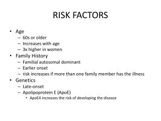
In a multivariate regression analysis that included several risk factors and measures of cardiovascular diseases (CVD), they found IMT of internal carotid artery to be the best predictor of transient ischemic attack (TIA) and stroke. The association seen between IMT of internal carotid artery and Magnetic Resonance Image (MRI) infarcts in TIA subjects in this study further indicates that the association also applies to morphological brain changes. Whether their presence suggests an increased risk for incident stroke is a matter that needs to be addressed in the future by long-term follow-up studies (Polak et al 1993). Cerebral lesions are generally considered to be the consequences of atherosclerosis, arteriosclerosis, cerebral hypoperfusion, or ischemia (Awad et al 1986, van Swieten et al 1991, Forsting et al 1991, Meyer et al 1992 and Price et al 1997). These cerebral lesions may be the precursors of clinical stroke (Forsting et al 1991, van Swieten et al 1991, Breteler et al 1994, Liao et al 1997 and Price et al 1997). 1. 3. Risk factors: Atherosclerosis is a disease of the old age group, although it has been reported in infants on rare occasions (Strong et al 1978). It is much commoner in men than in women up to the age of menopause; after that, the incidence tends to be at the same level in both sexes (Dawber 1980). Patients with high blood cholesterol tend to develop atheroma at an earlier age and to a more severe degree than do healthy subjects as in diabetes mellitus, myxedema, nephrosis and xanthomatosis (Stamler 1978). This is an independent risk factor, but there are some other risk factors that accelerate the process of atherosclerosis especially when lipid abnormality is also present which are called the additive risk factors. These are age (Salonen and Salonen 1991), male sex (O'Leary et al 1991), hypertension (Psaty et all 1992.I and Bots et al 1993), and its medication (Psaty et al 1992.A and Thrift et al 1996), diabetes mellitus R B E H
(Bonithon-Kopp et al 1991 and Golden et al 2002), severe obesity (Abbott et al 1987, Blankenhorn et al 1993, Furberg et al 1994 and Crouse et al 1995), cigarette smoking (Hays et al 1996 and Villablanca et al 2000), alcohol consumption (Ding et al 2004), periodontitis (Beck et al 2001) and hypothyroidism (Hak et al.2000). Several studies have shown that hypertension, hyperlipidemia, male sex, age, smoking, and postmenopausal status are consistent and independent factors that contribute to increasing carotid IMT (Heiss et al 1991, O'Leary et al 1992 and Wagenknecht et al 1997). The prevalence and severity of the disease and therefore the age when it is likely to cause tissue or organ injury are related to a number of factors, some constitutional and therefore immutable, but others acquired and potentially controllable. The constitutional factors include age, sex and familial background (Stamler 1978). There are four major acquired risk factors that are at least in some part amenable to control. They are hyperlipidemia, hypertension, diabetes mellitus and cigarette smoking. In addition there are a number of less important "soft" risks. These minor factors are associated with a less pronounced and difficult-to-quantitate risk. These include insufficient regular physical activity, competitive stressful life style, obesity, oral contraceptives, hyperuricemia, high carbohydrate intake and hyperhomocysteinemia (Stamler 1978). Multiple factors had been shown to impose more than additive effect. When three risk factors are present, e.g., hyperlipidemia, hypertension and smoking, the heart attack rate is seven greater than when none are present. Two risk factors increase the risk fourfold. However, the converse is equally important i.e., atherosclerosis may develop in absence of any apparent risk factors (McSween and Whaley 1992). R B E H
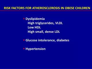
1. 3. 1. Hyperlipidemia: Hyperlipidemia is universally acknowledged to be a major factor for atherosclerosis (Oalmann et al 1981). Most of the evidence implicates hypercholesterolemia (Haust 1978), but hypertriglyceridemia may also play a role, although it is not as significant as hypercholesterolemia (Gotto 1979). The major evidence implicating hypercholesterolemia in the genesis of atherosclerosis includes the following: High cholesterol diets can produce Atherosclerotic plaques in experimental animals, including nonhuman primates, which are nearly identical to those, observed in human disease (Verschuren et al 2005). The major lipids in atheromas (plaques) are cholesterol and cholesteryl esters derived from the plasma (Grundy et al 2004). Many large-scale epidemiological analyses have demonstrated a significant correlation between the total plasma cholesterol or LDL level and the severity of atherosclerosis as judged by the mortality rate from ischemic heart disease (Boon et al 1994). The higher the total cholesterol level, the greater the symptomatic and fatal atherosclerotic disease. No threshold clearly separates persons at risk from those free of risk, but in general, atherosclerotic events are very uncommon with total cholesterol levels below 150 mg/dl. Hypertriglyceridemia, as manifested by elevated very low density lipoproteins (VLDL) levels, is also associated with some increased risk (McSween and Whaley 1992). Genetic or acquired disorders (e.g., familial hypercholesterolemia, diabetes mellitus and Hypothyroidism) that cause hypercholesterolemia lead to premature and severe atherosclerosis; an example is familial hypercholesterolemia, which in the homozygous state is often associated with myocardial infarction before age 20 years (Wendelhag et al 1992). When levels of serum cholesterol are lowered by diet or drugs (Blankenhorn et al 1993 and Chen and Farese 2004), there is evidence in animals that some of atherosclerotic plaques regress, or fail to progress within months and that the risk of cardiovascular mortality in selected patients is reduced. R B E H
Genetic manipulations of lipid components in mice (e.g. apolipoprotein E deficiency) lead to abnormal lipid metabolism and atheromatous lesions in these animals (McSween and Whaley 1992). It was also found that apolipoprotein E promotes the regression of atherosclerosis independently of lowering plasma cholesterol levels (Raffai et al 2005). The essential factor associated with development of atherosclerosis is elevated serum total cholesterol or low density lipoproteins (LDL), low high density lipoproteins (HDL) or both (Wendelhag et al 1992). High density lipoproteins and were found to be protective against stroke, whereas lipoprotein-a increased the risk (Jeng et al 1999 and Sacco et al 2001). When other potential factors are taken into account, including hypertension, diabetes, coronary heart disease (CHD), smoking, body mass and socio-economic factors (education); individuals with elevated HDL levels benefited from a reduction in the risk for stroke. People with HDL in the range of 35–50mg/dl demonstrated a lower risk. This protective effect was even greater in those with a HDL above 50 mg/dl. A 5mg/dl increase in HDL resulted in approximately a 24% reduction in stroke risk (McSween and Whaley 1992). If stroke is subdivided into atherosclerotic (large carotid artery disease, intracranial atherosclerotic disease) and non-atherosclerotic (cryptogenic, lacunar and cardio-embolic strokes) categories; the protective effect of HDL is increased still further in events of atherosclerotic origin. Greater protection with intermediate and high HDL levels was seen in the atherosclerotic compared with the non-atherosclerotic subgroup. This suggests that the effect of HDL may be greater in the atherosclerotic stroke subgroup. This is in line with the cardioprotective effect seen in coronary heart disease (Sacco 2001). It is important at this point to emphasize the inverse relationship between symptomatic atherosclerosis and the HDL level. High density lipoprotein R B E H
participates in reverse transport of cholesterol and is believed to mobilize this lipid from cells and presumably from atherosclerotic plaques and transport it to the liver for excretion in the bile. The higher the levels of HDL, the lower are the risk of ischemic heart disease. Hence, there is a great interest in dietary methods of lowering serum LDL and raising serum HDL. Nondietary influences may also affect the level of blood lipids. Exercise and moderate consumption of ethanol both raise the HDL level, whereas obesity and smoking lower it (McSween and Whaley 1992). 3. 2. Hypertension: The term hypertension, used without qualification, is synonymous with systemic arterial hypertension. However, hypertension is defined as raised pressure in a vascular bed. It affects 15-20% of population in many developed countries (Kashgarian 1985). Blood pressure rises through childhood and adolescence and reaches the plateau of normal adult levels in the third decade. However, mean blood pressure continues to increase with age but there is considerable individual variation in this increase and any diving line between normal and abnormal is arbitrary (Berglund 1989). Rough working definitions of hypertension have been laid down by the World Health Organization as follows: Hypertension: systolic pressure ≥160mmHg and/or diastolic pressure ≥95mmHg. Border line hypertension: systolic pressure =140-160 mmHg and/or diastolic pressure =90–95mmHg (McSween and Whaley 1992 and Kaplan 1997). R B E H
In about 95% of cases of hypertension the cause is not apparent and these patients are said to have primary, essential or idiopathic hypertension (which is either benign 90% or malignant 10%). In the remaining 5% hypertension is secondary to other disease processes. Benign hypertension causes changes in arteries of all sizes. In vessels from the aorta down to the smallest arteries, the changes are widespread and are termed hypertensive arteriosclerosis. The arteriosclerotic changes are similar to those observed in normotensive elderly subjects (Castleman and Smithwick 1948, Kannel, Schwartz and McNamara 1969 and Berk and Alexander 1996). In prospective studies increased blood pressure (both systolic and diastolic) has been consistently shown to be associated with a subsequent increased risk of ischemic heart diseases. In some community studies the risk in the 20% of the population with the highest pressures was four times that for the 20% with the lowest pressures. The relationship between blood pressure and ischemic heart diseases is not a simple linear one and there is considerable clinical debate as to the levels above which the risk is increased (Braunwald 1991). Hypertension is a major risk factor for atherosclerosis at all ages and, after age 45, may well be more important than hypercholesterolemia (Kannel et al 1970.L). Men age 45-62 years whose blood pressure exceeds 150/95mmHg have a more than fivefold greater risk of IHD than those with blood pressures of 140/90mmHg or lower. Both systolic and diastolic levels are important in increasing risk (Kannel et al 1976 and Zanchetti 1997). Hypertension has long been known to be a major risk factor for stroke (Kannel et al 1970, D'Agostino et al 1994 and Chanorro et al 1996) with systolic pressure appearing to be a stronger risk factor than diastolic (Rutan et al 1988). R B E H
Bhadelia et al in 1999, however, found that transient ischemic attack (TIA) subjects with MRI infarcts 3 mm in maximum diameter have significantly higher diastolic blood pressure and carotid wall IMT than TIA subjects without infarcts. This relationship was independent of age, sex, and other risk factors. Moreover, increasing values of diastolic blood pressure and internal carotid artery IMT were associated with higher risk of MRI infarcts in subjects with TIA. Association between diastolic blood pressure and brain infarcts has been demonstrated before by imaging and autopsy studies. This relationship between diastolic blood pressure and brain infarcts is believed to be due to hypertension-induced increase in cerebral microvascular tone (Mast et al 1995, Shinkawa et al 1995 and Chanorro et al 1996). 3. 3. Smoking: Endothelial dysfunction, altered lipid metabolism and adrenergic stimulation induced by smoking can lead to vascular damage (Hays et al 1996 and Villablanca et al 2000). Smoking is associated with increased risk for cardiovascular disease (Stamler et al 1993). A prospective study by Kannel and his group clearly showed that smokers had a threefold higher rate of sudden death (concerning heart disease) than did non-smokers (Kannel et al 1975). Smoking is thought to account for the relatively recent increase in the incidence and severity of atherosclerosis in women. When one or more packs of cigarettes are smoked per day for several years, the death rate from IHD increases up by 200%. As with serum cholesterol and hypertension, the risk is continuous. The person smoking more has a greater risk of having a major coronary event than a person who smokes less. Cessation of smoking reduces this increased risk in time (Strong and Richards 1976). R B E H
Cigarette smoking was found to be one of the major contributing factors to the pathogenesis of atherosclerosis (McGill et al 1963). In 1962, the Albany and Farmingham studies clearly demonstrated the definite relationship between cigarette smoking and morbidity and mortality from myocardial infarction and death from coronary disease (Doyle et al 1962). Other studies by Paul et al 1963 and Kannel et al 1966 added weight to the theory that cigarettes played a major role in the formation of atherosclerosis. A long term study by Paffenbarger et al in 1966indicated that the smoking habits of college students (dating back as far as 1926) were statistically related to their development of coronary disease in later life. In 1964, the Surgeon General's report indicated that cigarette smokers (greater than one pack per day) had a three times greater chance of having a coronary event than non-smokers. Epidemiological studies, therefore, have been convincingly relating cigarette smoking to the development of atherosclerosis. There has been equally good evidence from the autopsy table correlating the amount of smoking and coronary atherosclerosis since when Auerbach and his associates studied the degree of atherosclerosis in 1372 men who died of disease other than coronary heart disease. Experienced interviewers (who were not familiar with the autopsy findings) obtained histories from the families of these men concerning their past smoking habits. This study showed not only that smokers have higher percentage of coronary atherosclerosis than non-smokers, but that "within age group, the proportion of cigarette smokers with an advanced degree of atherosclerosis increased constantly with the amount of cigarette smoking " (Auerbach et al 1965). R B E H
In animal experiments, nicotine has been administered in large amounts without atherogenic effects except for the suggestion that it may cause necrosis and calcifications of the medial arterial layers (Hammond 1966 and Kahn 1966). Coronary arteriography studies have demonstrated a correlation between the number of cigarettes consumed and the severity of coronary artery disease as well as the accelerating effect of cigarette consumption on the development of coronary artery disease (Herbert 1975). It has been though for years that nicotine causes vasoconstriction as well as increased heart rate and blood pressure secondary to discharge of catecholamines. However, evidence from the experiments of Cryer et al 1976 strongly suggests that the hemodynamic effects of smoking are not mediated by the elevated plasma chatecholamines but are the result of immediate local release of norepinephrine from sympathetic nerves innervating the heart and other tissues. This study showed that within 10 minutes of the start of smoking there was a significant elevation of plasma norepinephrine and epinephrine. The increase in pulse rate and blood pressure preceded the elevations of plasma catecholamines and was so prevented by prior adrenergic blockade. Aronow in 1976 had centered a great deal of investigation around the effects of carbon monoxide on the cardiovascular system. It is known that smokers have CO levels of 400ppm in their pulmonary capillary blood which causes high carboxyhemoglobin (COHb) levels (10-18%). Aronow has also shown that since the "affinity of hemoglobin for CO is approximately 245 times greater than its affinity for oxygen; CO displaces oxygen from hemoglobin, reducing the amount of oxygen available to the myocardium". Carbon monoxide causes a shift to the left of the oxygen-hemoglobin (O2- Hb) dissociation curve, which in turn causes a tighter binding of O2 to Hb and thereby decreases the availability of O2 to the arterial wall and myocardium (Ayres et al 1970). R B E H
Hypoxia alone (10% O2) in cholesterol-fed rabbits produces lesions in the arterial wall indistinguishable from those found in similarly fed rabbits with COHb levels of 15% (Astrup et al 1968and Kjeldsen et al 1968). The aortas, femoral and coronary vessels all showed lipid deposition of a higher degree in the experimental CO-exposed rabbits than the control rabbits that were fed only cholesterol; the average content of cholesterol per 100g wet weight of aortic tissue was 703mg in the control group and 1774mg in the CO-exposed group. Rabbits on a normal diet and exposed to CO for 13 weeks with levels of COHb of 10%-11% developed focal intimal and subintimal changes in a significantly higher degree than nonexposed control animals (Astrup et al 1968). Triggering lipid accumulation in the arterial lumen (Whereat et al 1970) and increasing platelet stickiness (Birnstingl et al 1971) are two other biochemical effects of carbon monoxide. It can be concluded that cigarette smoking has a definite effect on the formation of atherosclerosis, and more specifically, that carbon monoxide can be a major factor in the acceleration of the atherosclerotic process. 1. 3. 4. Diabetes mellitus: Diabetes mellitus is not a single disease but the pathological and metabolic state caused by inadequate insulin action. A feature common to all types is glucose intolerance. It is defined clinically as either a fasting plasma glucose level greater than 140mg/dl or a two hour post-prandial plasma glucose greater than 200mg/dl (McSween and Whaley 1992). R B E H
It has been recognized for years that the main problem in diabetes mellitus is the enhanced risk of premature atherosclerosis. This has been documented extensively by retrospective and autopsy studies (Kissane 1990). Insulin is a major anabolic hormone. In addition to its other functions, it promotes the uptake of free fatty acids by adipose tissue. Insulin lack therefore, results in general catabolic state hyperlipidemia due to lypolysis in adipose tissue (Volk and Arquilla 1985 and Kawachi 2004). Diabetes mellitus is classified into type1 (insulin dependent) and type2 (insulin independent). Type2 diabetes mellitus is ten times more common than type1 and it affects obese subjects over 40 years (Cudworth 1978). Selective destruction of insulin secreting B-cells in pancreas occurs in type1 diabetes mellitus resulting in insulitis with evidence that genetic factors, autoimmunity and possibly viral infection may all be etiologically involved (Foulis 1987). In type2 diabetes mellitus there may be both qualitative and quantitative insulin insufficiency due to resistance to the action of insulin. These patients have to hypersecrete insulin to achieve metabolic homeostasis with resultant B-cells exhaustion (Cerasi and Luft 1967). Diabetic macroangiopathy is most commonly affecting large muscular arteries and microangiopathy affecting arterioles and capillaries (Williamson and Kilo 1977). Macroangiopathy is simply atheroma, which tends to develop early and become severe in diabetics of either sex. This, plus the fact that 50% of patients with type2 diabetes mellitus have hypertension, results in 80% of adult diabetic deaths being due to cardiovascular, cerebrovascular or peripheral vascular diseases together with an increased susceptibility to bacterial infection (Volk and Arquilla 1985). R B E H
Impaired glucose tolerance is common in elderly subjects and has been demonstrated to be associated with increased prevalence of cardiovascular disease and its risk factors (Savage et al 1991). Prospective studies indicate that persons with atherosclerotic disease exhibit abnormalities in glucose tolerance more frequently than do clinical controls (Volk and Arquilla 1985). The Cardiovascular Health Study data confirm prior evidence that asymptomatic hyperglycemia is not a benign condition in the elderly and that its previously demonstrated association with coronary disease (Mykkanen et al 1992) also extends to cerebrovascular disease (Manolio et al 1996). Diabetes mellitus has shown strong and consistent relationships with stroke incidence, (Wolf et al 1977, Aronow et al 1988 and Manolio et al 1996). Less consistent associations have been demonstrated for impaired glucose tolerance, (Fuller et al 1983, Burchfiel et al 1994). Diabetes mellitus induces hypercholesterolemia and a markedly increased predisposition to atherosclerosis and when other factors are equal, the incidence of myocardial infarction is twice as high in diabetics as in non-diabetics. There is also an increased risk of stroke and, even more striking, perhaps a hundredfold increased risk of atherosclerosis-induced gangrene of the lower extremities. Indeed, in the absence of diabetes mellitus, atherosclerotic gangrene of the lower extremities is uncommon (Nerup et al 1987). Additional studies have confirmed that asymptomatic hyperglycemia is also a significant risk factor for coronary heart disease (Epstein 1967). R B E H
There is evidence to suggest that some individuals respond to carbohydrate feeding with a rise in serum lipids. Refined carbohydrates (used in cookies, candies, pies) are especially suspect as lipogenic factors which could enhance the serum lipid rise cholesterol deposition (Gotto et al 1980). 3. 5. Age: Age is one of the additive risk factors of atherosclerosis (Campbell et al 1989 and Salonen and Salonen 1993). Stroke incidence rises sharply with advancing age in middle age (Kagan et al 1980, Wolf et al 1987 and Davis et al 1987) and in the elderly (Wolf et al 1987, Welin et al 1987, Woo and Lau 1990 and Zeiler et al 1992). Vermeer et al in 2003 found that the incidence of silent brain infarcts on MRI in the general elderly population strongly increases with age. Other authors found that cerebral abnormalities, such as infarctions and white matter lesions are frequently detected in older individuals (Awad et al 1986, George et al 1986, Meyer et al 1992, Boone et al 1992, Bots et al 1993 and Price et al 1997). 3. 6. Gender: Male sex was found to be an additive risk factor of atherosclerosis (McGill et al 1977, Bonithon-Kopp et al 1991 and O'Leary et al 1991) and stroke incidence was more than three times higher in women aged 80 years and older than in women aged 65-74 and nearly twice as high in men aged 80 years and older compared with those aged 65-74, but stroke incidence did not differ by sex in the full age range although there was a R B E H
borderline significant trend toward greater incidence in men aged 65-74 years than women of the same age (Manolio et al 1996). Gender relationships with stroke incidence show men at higher (Kannel et al 1983), similar (Manolio and Furberg 1992) or lower (D'Alessandro et al 1992) risk than women. The significant association of gender with stroke after excluding subclinical disease measures suggests that any sex differences in stroke incidence in these data are related to differences in subclinical disease between women and men (Manolio et al 1996). 1. 3. 7. Periodontitis: Several studies have suggested that periodontitis is also associated with coronary heart disease and cerebrovascular disease (Syrajanen et al 1989, Mattila et al 1989, DeStefano et al 1993, Mattila et al 1993, Kweider et al 1993, Mattila et al 1995, Beck et al 1996, Joshipura et al 1996, Grau et al 1997, Loesche et al 1998 and Morrison et al 1999). A mechanism has been proposed whereby periodontitis creates a burden of bacterial pathogens, antigens, endotoxins and inflammatory cytokines that contribute to the process of atherogenesis and thromboembolic events. Certain persons may exhibit greater expression of local and systemic mediators in response to infection and inflammation and may thereby be at increased risk for atherosclerosis. The atherosclerosis process may result in decreased arterial patency and/or decreased compliance of the vessel. Ultimately, atherosclerotic lesions may fissure and/or rupture, resulting in occlusion of the vessel lumen, precipitating a myocardial infarction or stroke (Beck et al 2001). R B E H
Elkind et al in 1999 found that individuals with periodontal disease (abnormal probing depth >3mm, significant attachment loss >3.5mm, or with no teeth) had greater atherosclerotic plaque thickness. Measures of both current and cumulative periodontitis became more severe as tooth loss increased. A significant association was observed between tooth loss levels and carotid artery plaque prevalence. Among those with 0-9 missing teeth, 46% had carotid artery plaque, whereas among those with 10 missing teeth, carotid artery plaque prevalence was 60%. These data suggest that tooth loss is a marker of past periodontal disease and is related to subclinical atherosclerosis, thereby providing a potential pathway for a relationship with clinical events (Desvarieux et al 2003). 1.3. 8. Hypothyroidism: Another risk factor is overt hypothyroidism which has been found to be associated with cardiovascular disease. Atherosclerosis occurs in the hypothyroid patient as a result of angiotensin produced arterial constriction with its resultant damage to the intimal lining of the arteries, at which sites cholesterol is deposited. Untreated, the cholesterol deposits increase to be eventually replaced by calcium. When treated appropriately early enough, the cholesterol deposits can be reabsorbed (Alford 2000 and Hak et al 2000). 1.3. 9. Aspirin intake: The regular use of aspirin was found to reduce the risk of ischemic stroke for many patients with clinically manifest atherosclerotic vascular R B E H
disease (Antiplatelet Trialists Collaboration 1994). In contrast, randomized clinical trials involving people at relatively low risk for stroke have shown an opposite trend. Although aggregate data are inconclusive, these trials associated aspirin use with an increased risk of stroke (Peto et al 1988, Steering Committee of the Physicians' Health Study Research Group 1989, and Early Treatment of Diabetic Retinopathy Study Investigators 1992). In a previously reported analysis of the population-based Cardiovascular Health Study, regular aspirin use emerged as an independent risk factor for stroke even after adjustment for other stroke risk factors (Manolio et al 1996). 1. 3. 10. Alcohol consumption: Alcohol consumption is a controversial risk factor, excessive alcohol intake although it is clear that it is deleterious, moderate consumption appears to protect against stroke (Sacco et al 1999). It became clear that individuals who ingest 5 or more units of alcohol per day had a significantly increased risk for ischemic stroke. Importantly, those who kept to 1 or 2 units per day had a lower ischemic stroke risk than those who did not drink at all (Sacco 2001). These findings support those of other studies, and the recommendation made in the National Stroke Association Stroke Prevention Guidelines is that drinking in moderation is beneficial for those who drink alcohol and have no health contraindications to alcohol use. People who do not drink should not be encouraged to do so; however, those who do should be encouraged to drink the appropriate amount (Gorelick et al 1999). R B E H