Brain growth and development
Brain growth and development. Growth Spurt. % of increase of growth.

Brain growth and development
E N D
Presentation Transcript
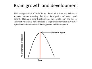
Brain growth and development Growth Spurt % of increase of growth The weight curve of brain is not linear with time but follows a sigmoid pattern meaning that there is a period of more rapid growth. This rapid growth is known as the growth spurt and this is the most vulnerable period where a slightest disturbance may have a profound effect on overall brain growth and development. Time
Species difference: In some species Growth spurt occurs prenatally (guinea pig, Sheep, Monkey) whereas it occurs postnatally in other sp. (Human, Rat, Rabbit )
Growth of the Brain • Development (hypertrophy) • Cells are developed into mature functioning units as a result of deposition of different cellular constituents (protein, lipid). • Mature cells differ from immature ones in both size and chemical composition. • Growth (hyperplasia) • An increase in the no. of cells until the adult cellular population is achieved in the tissue. • Determined by the genetic make up of the individual organs. Brain growth and development can be divided into 4 phases: Phase 1: growth proceeds entirely by cell multiplication (Hyperplasia alone), with a proportional increase in weight, and in protein or DNA or lipid content. At the end of this phase DNA synthesis is slowed down , gradually but accumulation of other compounds continues. Phase 2: It is the combination of both hyperplasia and hyperthophy, in which there is an increase in cell size and smaller increase in cell number. The rate of DNA accretion slows down further, but the accumulation of other constituents continues. Phase 3: DNA synthesis is stopped altogether and cells developed by increasing in size and continued synthesis and accumulation of proteins and lipids. In this phase only hypertrophy persists. Phase 4: Construction of synaptic interneuronal junctions.
Events that occur in 3 Phases: Phase 1: Organogenesis and neuronal multiplication Phase 2: Axonal and dendritic growth ; Glial multiplication; Myelination Phase 3: Growth in size Phase 4: Synaptogenesis
Postnatal period Prenatal period
These final structures differentiate during the fetal period of brain development.
Brain Development The brain grows at an amazing rate during development. At times during brain development, 250,000 neurons are added every minute!! At birth, almost all the neurons that the brain will ever have are present. However, the brain continues to grow for a few years after birth. By the age of 2 years old, the brain is about 80% of the adult size. You may wonder, "How does the brain continue to grow, if the brain has most of the neurons it will get when you are born?". The answer is in glial cells. Glia continues to divide and multiply. Glia carries out many important functions for normal brain function including insulating nerve cells with myelin. The neurons in the brain also make many new connections after birth.
Multiplication of Neurons and Gilal cells are two consecutive process. The former being followed by the later. Neurogenesis: Neurons originate from the stem cells (Neuroblast) in the neuroepithelium. Gliogenesis: In vertebrates both neuro and gliogenesis occurs in neuroepithelium. Birth Rapid Gliogenesis DNA-Phosphate Slow Rapid Neurogenesis Weeks
Only recently has it become generally accepted that new neurons are added in discrete regions of the adult mammalian CNS. • Active neurogenesis occurs throughout the life in • Subventricular zone (SVZ) of the lateral ventricle • Subgranular zone (SGZ) of the dentate gyrus in the hippocampus • Other regions are non-neurogenic but neurogenesis can take place in this regions only after brain insults
Neurogenesis in the Subventricular Zone • The forebrain subventricular zone (SVZ) is a prolific source of • neuronal precursors (neural stem cells). • These neuronal progenitor cells (which give rise to neurons) migrate • to the olfactory bulb by means of a restricted pathway known as the • rostral migratory stream (RMS). • Once in the core of the olfactory bulb, the cells migrate in a radial pattern and differentiate into interneurons.
Cell Migration along the RMS Confocal microscopy images of a cell migrating along the RMS
Neural cell death Endogenous reasons: Some Neural cells are subjected to death during developmental process in CNS. Reasons: Lac of maintenance from target tissue. Target tissue generally release one or more neurotrophic factors such as Nerve growth factors (NGF) which are proved to be essential for the survival of neurons. In most cases neuronal cells die if they don’t receive adequate NGF from neuroepithelial cells. CNS also regulates the total no of neurons by stimulating neural cell death. During early periods of development many inaccurate and aberrant synaptic interconnections are formed in the nervous tissue which is removed by mediating neural cell death.
Stem cell therapy for neurodegeneration Many common neurological disorders, such as Parkinson’s disease, stroke and multiple sclerosis, are caused by a loss of neurons and glial cells. In recent years, neurons and glia have been generated successfully from stem cells in culture, fuelling efforts to develop stem-cell-based transplantation therapies for human patients. Stem cells would be isolated and transplanted to the diseased brain and spinal cord, either directly or after predifferentiation/genetic modification in culture to form specific types of neuron and glial cell, or cells producing neuroprotective molecules.
In strategies relying on stimulation of the patient's own repair mechanisms, endogenous stem cells would be recruited to areas of the adult brain and spinal cord affected by disease, where they would produce new neurons and glia (neurogenic and gliogenic areas along lateral ventricle and central canal are shown in hatched red). Stem cells could provide clinical benefits by neuronal replacement, remyelination and neuroprotection.
Role of microRNAs during brain development MicroRNA (miRNA) are a newly recognized class of small, noncoding RNA molecules that participate in the developmental control of gene expression. The regulation of a set of highly expressed neural miRNA during mouse and human brain development has been identified recently. Putative mRNA targets for developmentally regulated miRNAs Identifying the miRNA targets The miRNA and its putative target must be expressed in the same tissue. Putative targets may undergo posttranscriptional regulation that is coordinated with the miRNA expression. Combining this candidate strategy with a search of the NCBI databases revealed several genes with a high degree of complementarity to developmentally regulated neuronal miRNA
Calcineurin A, an isoform of the calcineurin catalytic subunit, fits these target criteria for miR- 131. Calcineurin is a major phosphatase of the central nervous system involved in a variety of neuronal signaling cascades, which plays a critical role in longterm depression. In humans and rodents, miR-131 demonstrates 19 nt and 18 nt, respectively, out of 21 nt complementarity to a sequence within the 3 UTR of calcineurin A mRNA. Calcineurin A, and particularly its expression in developing brain is regulated posttranscriptionally, most likely at the translational level
mRNA for Id2 (Inhibitor of DNA binding), the protein antagonizing neuronal differentiation, demonstrates 20 nt out of 23 nt complementarity to miR-9. So upregulation of this mir-9 expression occurs during development.
A sequence within DNA helicase SMBP2 transcript is nearly complementary to miR-103. Mutation of this protein causes mouse neuromuscular degeneration , a disease similar to human spinal muscular atrophy and linked to miRNA-containing complex. Otx1, murine homolog of the Drosophila transcription factor orthodenticle, is necessary for normal corticogenesis, and may represent another putative target for miR-103.