M-2 HEPATOBILIARY IMAGING
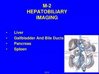
M-2 HEPATOBILIARY IMAGING. Liver Gallbladder And Bile Ducts Pancreas Spleen. 2014. HEPATIC ANATOMY. Catalano, O. A. et al. Radiographics 2008;28:359-378. GOALS. Review anatomy of hepato- biliary system. Correlate imaging with pathology.

M-2 HEPATOBILIARY IMAGING
E N D
Presentation Transcript
M-2HEPATOBILIARY IMAGING Liver Gallbladder And Bile Ducts Pancreas Spleen 2014
HEPATIC ANATOMY Catalano, O. A. et al. Radiographics 2008;28:359-378
GOALS • Review anatomy of hepato- biliary system. • Correlate imaging with pathology. • Discuss radiologic imaging options. • Choose treatment
ANATOMY / PHYSIOLOGY Portal vein flow Hepatic arterial flow Hepatic vein flow Biliary drainage
PORTAL BLOOD FLOW The Portal vein is formed by the juncture of the Splenic vein and the Superior Mesenteric vein. The Inferior Mesenteric vein usually joins the Splenic vein.
PORTAL VEIN Coronal and Axial images CT US
HEPATIC ARTERIAL FLOWLATERAL AORTOGRAM SHOWS ORIGIN OF CELIAC ARTERY AND THE SUPERIOR MESENTERIC ARTERY CELIAC The Celiac Artery splits into 3 branches: Supplies Diaphragmatic organs 1- Common Hepatic Artery. 2- Splenic Artery. 3- Lt. Gastric Artery. SMA
THE COMMON HEPATIC ARTERY BECOMES THE PROPER HEPATIC ARTERY AFTER THE GASTRODUODENAL BRANCH DESCENDS. Proper hepatic * Celiac Gastroduodenal SMA * The Lt. gastric is small and out of the section on this image
HEPATIC VEINS Coronal scan
HEPATIC VEINS ENTERING IVC CT ULTRASOUND
WHAT IS THE PRESENTATION OF HEPATIC VEIN THROMBOSIS?(Budd Chiari syndrome) • Enlarged edematous liver • Painful-capsular pressure • Ascites-pressure effects • Hypercoagulable states- etiology • Elevated liver enzymes- infarction • Diagnosed with Hepatic Venography • Or CT
NORMAL BILIARY ANATOMY ULTRASOUND GALLBLADDER COMMON BILE DUCT Silva, A. C. et al. Radiographics 2004;24:677-687
NORMAL BILIARY ANATOMY MR CHOLANGIOGRAM (MRCP) Silva, A. C. et al. Radiographics 2004;24:677-687
ENDOSCOPIC RETROGRADE Cholangio - Pancreatography ERCP MR cholangiogram shows signal from the bile and other fluids. ERCP has iodinated contrast injected with an endoscope with the canula in the distal common bile duct.
OPERATIVE CHOLANGIOGRAM HEPATO-BILIARY SCAN - HIDA
DARK GREEN EMESIS WOULD BE TYPICAL FOR GI OBSTRUCTION. • Pyloric stenosis • Duodenal atresia • Annular pancreas • Gallstone ileus
WHO PRESENTS FOR IMAGING? • Right upper quadrant pain • Altered laboratory data • Staging of malignancy / infection • Physical exam findings • Abdominal trauma
ACUTE RIGHT UPPER QUADRANT PAIN • Differential Diagnosis: • Acute Cholecystitis • PUD / Gastritis / Reflux • Acute Hepatitis • Pancreatitis
RIGHT UPPER QUADRANT PAIN • Gallstone = cholelithiasis- 10% prevalence • Stone impaction and obstruction cystic duct • Pain with contraction after fatty meal 20-30 minutes • Adult 40+- female more common
DIAGNOSIS ULTRASOUND • Cost / Availability • Fluid background is ideal for imaging • Helpful to assess for any associated biliary dilatation or inflammatory change
MURPHY’S SIGN A Sonographic Murphy’s sign is focal tenderness corresponding to the gallbladder. Along with other ultrasound evidence of inflammation (gallbladder wall thickening, pericholecystic fluid) it helps physicians separate Acute Cholecystitis from gallstones alone.
CHOLECYSTITISWith diffuse wall thickening and edema. Ultrasound and CT demostration of edema in and around GB wall
CHOLECYSTITIS QUESTION? Pain is often referred to other location with cholecystitis, Which is the correct answer? 1--Shoulder 2--Umbilicus 3--EG junction 4--Back
IMAGING ALTERNATIVES • Nuclear medicine - HIDA • CT • X-ray • Cholangiography - MR or Endoscopic
HEPATO - BILIARY SCINTIGRAM NORMAL HIDA Gall bladder Obstructed cystic duct doesn’t allow for filling of radionuclide into the GB. Absent Gall bladder ABNORMAL HIDA
GALLSTONES 15-30% calcify
COMPLICATIONS OF GALLSTONES Cystic duct obstruction Cholecystitis A Common bile duct obstruction Obstructive jaundice B Ascending cholangitis Pancreatic duct obstruction Pancreatitis C A B C
ALTERED LABORATORY DATA • Bilirubin - jaundice • Amylase / lipase - pancreatitis
QUESTION? HOW CAN A HEMOLYTIC ANEMIA (SICKLE CELL ANEMIA) GIVE AN ELEVATED DIRECT BILIRUBIN? 1--Hemolytic crisis 2--Transfusion Hepatitis 3--Choledocholithiasis 4--Hepatic infarction
CBD Obstructed duct due to distal calculus PV Normal bile duct size Diameter < portal diameter Note dilated CBD with impacted calculus
*Note dilated bile ducts. (Low density branching structures anterior to portal veins) Normal The Portal vein is opacified (white) from IV contrast administration. The biliary tree is of lower density and shows as a branching structure anterior to the portal vein.
Dilated CBD with calculi Normal size CBD Endoscopic retrograde Cholangiopancreatography ERCP
PANCREATITIS elevatedAMYLASE & LIPASE Biliary calculi-obstruction Alcohol- chemical toxicity
ACUTE PANCREATITISEDEMATOUS There is diffuse edema in and adjacent tissues around the pancreas.
A patient with diagnosis of pancreatitis who had developed a pseudocyst over past month Comes to the hospital with worsened pain and a blood pressure of 80/60. 1-Mesenteric arterial infarction 2-Portal vein thrombosis 3-Perforated ulcer 4-Leaking pseudoaneurysm
Normal vasculature Pseudo aneurysm
COMPLICATIONS OF PANCREATITIS • Pseudocyst • Pain • Infection • Hemorrhage- pseudoaneurysm • Pancreatic insufficiency Large retrogastric fluid collection is a pseudocyst related to pancreatic enzyme break down of tissue.
SPECIAL CASES • Emphysematous cholecystitis • Acalculous cholecystitis • Gallstone ileus
A 64-year-old man with insulin-dependent adult-onset diabetes mellitus seeks emergency medical treatment after 2 days of increasingly severe abdominal pain in the right upper quadrant that has spread over the entire abdomen and is associates with nausea, vomiting, fever and chills. On examination, he is alert and oriented but appears to be quite acutely distressed. Vital signs are temperature 39.4C (103F), pulse 140 beats per minute, and blood pressure 100/60mmHg. His sclerae are mildly icteric. His abdomen is diffusely tender with marked guarding in the right upper quadrant.
QUESTION? IN GALLSTONE ILEUS OBSTUCTION OF THE GI TRACT OCCURS COMMONLY IN THE: 1--Pyloric channel 2--Duodenal C loop 3--Ileocecal valve 4--Hepatic flexure
GALLSTONE ILEUS Small Bowel Obstruction at IC valve due to migration of gallstones that erode into duodenum from GB. 2002 1999
ACALCULOUS CHOLECYSTITISBILIARY STASIS - FASTING / ICU PATIENTS
SAME PATIENT ABDOMEN SCAN DONE 2/25/08